. 2025 Jun 3;11(6):431.
doi: 10.3390/gels11060431.

Nanoemulsion Hydrogel Delivery System of Hypericum perforatum L.: In Silico Design, In Vitro Antimicrobial-Toxicological Profiling, and In Vivo Wound-Healing Evaluation

Affiliations

Nanoemulsion Hydrogel Delivery System of Hypericum perforatum L.: In Silico Design, In Vitro Antimicrobial-Toxicological Profiling, and In Vivo Wound-Healing Evaluation

Ahmet Arif Kurt et al. Gels. .

Abstract

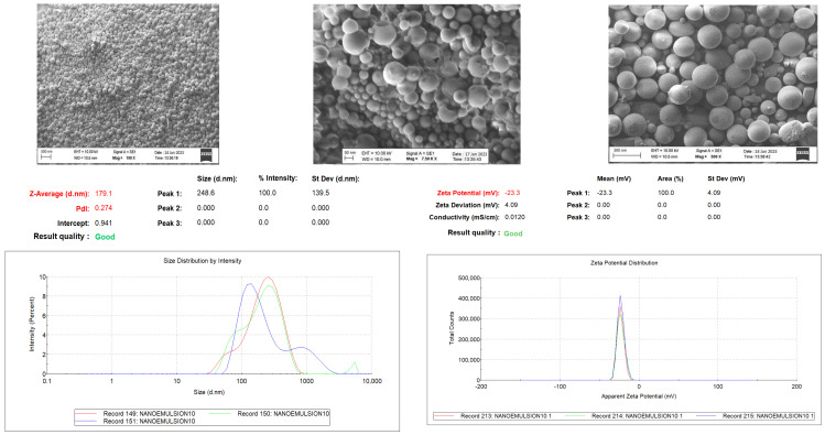
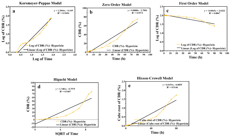
Hypericum perforatum L. (H.P.), a plant renowned for its wound-healing properties, was investigated for antioxidant/antimicrobial efficacy, toxicological safety, and in vivo wound-healing effects in this research to develop and characterize novel nanoemulsion hydrogel (NG) formulations. NG were prepared via emulsion diffusion-solvent evaporation and polymer hydration using Cremophor RH40 and Ultrez 21/30. A D-optimal design optimized oil/surfactant ratios, considering particle size, PDI, and drug loading. Antioxidant activity was tested via DPPH, ABTS+, and FRAP. Toxicological assessment followed HET-CAM (ICH-endorsed) and ICCVAM guidelines. The optimized NG-2 (NE-HPM-10 + U30 0.5%) demonstrated stable and pseudoplastic flow, with a particle size of 174.8 nm, PDI of 0.274, zeta potential of -23.3 mV, and 99.83% drug loading. Release followed the Korsmeyer-Peppas model. H.P. macerates/NEs showed potent antioxidant activity (DPPH IC50: 28.4 µg/mL; FRAP: 1.8 mmol, Fe2+/g: 0.3703 ± 0.041 mM TE/g). Antimicrobial effects against methicillin-resistant S. aureus (MIC: 12.5 µg/mL) and E. coli (MIC: 25 µg/mL) were significant. Stability studies showed no degradation. HET-CAM tests confirmed biocompatibility. Histopathology revealed accelerated re-epithelialization/collagen synthesis, with upregulated TGF-β1. The NG-2 formulation demonstrated robust antioxidant, antimicrobial, and wound-healing efficacy. Enhanced antibacterial activity and biocompatibility highlight its therapeutic potential. Clinical/pathological evaluations validated tissue regeneration without adverse effects, positioning H.P.-based nanoemulsions as promising for advanced wound care.

Keywords: Cremophor RH40; HET-CAM; Hypericum perforatum L.; antimicrobial activity; antioxidant activity; drug delivery systems; hydrogel; in silico modeling; nanoemulsion; wound healing.


Conflict of interest statement

The authors declare no conflicts of interest.

Figures

Figure 1
Figure 1
Three-dimensional (3 D) response surface contour plots were generated to visualize the relationships between the independent variables (A-Hypericum perforatum L. macerate and B-surfactant concentration (Cremophor RH-40)) and the dependent variables (particle size, particle size distribution, and zeta potential) based on the quadratic calibration model employed in the in-silico modeling (DoE-Design Expert 13).
Figure 2
Figure 2
SEM images, particle size distribution, and zeta potential graphs of NE-HPM10 (n = 3).
Figure 3
Figure 3
Viscosity, shear stress, and shear rate graphs of NG-1 (containing Ultrez 21) and NG-2 (containing Ultrez 30).
Figure 4
Figure 4
Release kinetic graphs of NG-2 formulation: (a) Korsmeyer–Peppas; (b) zero order; (c) first order; (d) Higuchi; (e) Hixson–Crowell kinetic models. CDR: cumulative drug release quantity, SQRTT: square root of time.
Figure 5
Figure 5
Graph of % cumulative hypericin content versus time.
Figure 6
Figure 6
Minimum inhibitory (MIC) and minimum bactericidal/fungicidal (MBC/MFC) concentrations of different antimicrobial agents. Groups: 1–olive oil, 2—HPM (H. perforatum L. macerate), 3—NE-HPM-10, 4—CP1, 5–6—CP2, 7—CP3, 8—negative control (S.F.), 9—positive control (Sefalosporin).
Figure 7
Figure 7
DPPH, ABTS, and FRAP antioxidant activity results.
Figure 8
Figure 8
HET-CAM images of all groups (1: HPM; 2: NE-10 Placebo; 3: NE-HPM-10; 4: CP1; 5: CP2; 6: CP:3).
Figure 9
Figure 9
Wound-healing score of Group A (HPM), Group B (NG-2), Group C (CP1), Group D (CP2), Group E (CP3), and Group F (Control) in the skin defect area after 4, 8, and 12 days.
Figure 10
Figure 10
Wound-healing images of Group A (HPM), Group B (NG-2), Group C (CP1), Group D (CP2), Group E (CP3), and Group F (Control) in the skin defect area after 4, 8, and 12 days.
Figure 11
Figure 11
Histopathological appearance of wound healing in the skin defect area according to groups. (A) Group CP1 exhibited incomplete epithelialization and mild connective tissue proliferation; (B) group Hypericum perforatum macerate (HPM) showed initial epithelialization at the wound edges and increased connective tissue proliferation; (C) group nanoemulsion-Hypericum perforatum macerate + hydrogel (NG-2) displayed a significant increase in both epithelialization and connective tissue development; (D) group CP2 exhibited slight epithelial tissue closure and significant connective tissue healing; (E) group CP3 demonstrated modest yet notable epithelial tissue closure and substantial connective tissue healing; and (F) the control group showed minimal healing in both connective tissue and epithelialization, with the defect remaining patent. Arrows indicate the defect area (hematoxylin and eosin staining; scale bars = 200 µm).
Figure 12
Figure 12
Immunohistochemical staining revealed the following patterns of cytokeratin expression across the treatment groups. (A) Group CP1 exhibited mild cytokeratin expression; (B) group Hypericum perforatum macerate (HPM) showed increased cytokeratin expression at the wound margins; (C) group nanoemulsion-Hypericum perforatum macerate + hydrogel (NG-2) displayed a significant increase in cytokeratin expression; (D) group CP2 exhibited mild cytokeratin expression in epithelial cells; (E) group CP3 demonstrated a moderate increase in cytokeratin expression; and (F) the control group showed minimal cytokeratin expression in only a few cells of the epithelial layer. Arrows indicate immunopositive cells (streptavidin-biotin peroxidase method; scale bars = 50 µm).
Figure 13
Figure 13
Immunohistochemical staining revealed the following patterns of VEGF expression across the treatment groups. (A) Group CP1 exhibited minimal VEGF expression; (B) group Hypericum perforatum macerate (HPM) showed a modest increase in VEGF expression, particularly within epithelial cells; (C) group nanoemulsion-Hypericum perforatum macerate + hydrogel (NG-2) displayed a significant increase in VEGF expression; (D) group CP2 exhibited mild VEGF expression in epithelial cells; (E) group CP3 demonstrated a moderate increase in VEGF expression within epithelial cells; and (F) the control group showed very faint VEGF expression in only a few cells. Arrows indicate immunopositive cells (streptavidin-biotin peroxidase method; scale bars = 50 µm).
Figure 14
Figure 14
Appearance of Type III and Type I collagen formation according to groups. (A) Group CP1 showing very prominent Type III collagen (green) and very slight Type I collagen (red) (arrow), (B) group Hypericum perforatum macerate (H.P.M.) showing significant Type III collagen and slight Type I collagen (arrow), (C) group nanoemulsion form-Hypericum perforatum macerate + hydrogel (NG-2) showing a significant decrease in Type III and an increase in Type I collagen (arrow), (D) group CP2 showing a significant decrease in Type III and an increase in Type I collagen (arrow), (E) group CP3 showing decreased Type III and increased Type I collagen (arrow), and (F) control group showing very prominent Type III collagen and very slight Type I collagen (arrow). Arrows indicate areas with red fluorescence, representing mature Type I collagen. Picro-Sirius Red method, bars = 50 µm.

Similar articles

References

    1. Wu J., Xue W., Yun Z., Liu Q., Sun X. Biomedical applications of stimuli-responsive “smart” interpenetrating polymer network hydrogels. Mater. Today Bio. 2024;25:100998. doi: 10.1016/j.mtbio.2024.100998. - DOI - PMC - PubMed
    1. Duman G., Aslan İ., Yekta Özer A., İnanç İ., Taralp A. Liposome, gel and lipogelosome formulations containing sodium hyaluronate. J. Liposome Res. 2014;24:259–269. doi: 10.3109/08982104.2014.907305. - DOI - PubMed
    1. Tavasli A., Navidfar A., Inangil D., Inangil G., Aslan I., Trabzon L. Hydrophilic sol-gel TiO2 coating on procaine-loaded injector needle for painless clinical treatments. Colloids Surf. A Physicochem. Eng. Asp. 2024;687:133556. doi: 10.1016/j.colsurfa.2024.133556. - DOI
    1. Hunt M., Torres M., Bachar-Wikstrom E., Wikstrom J.D. Cellular and molecular roles of reactive oxygen species in wound healing. Commun. Biol. 2024;7:1534. doi: 10.1038/s42003-024-07219-w. - DOI - PMC - PubMed
    1. Srivastava M.K. Advances in Mergers and Acquisitions. Volume 23. Emerald Publishing Limited; Bingley, UK: 2024. Strategic Choices Between Alliances and Acquisitions: What Do We Know and Where Do We Go from Here? pp. 123–131.

LinkOut - more resources