Review
. 2025 Jun 9;11(6):441.
doi: 10.3390/gels11060441.

Biomaterials in Postoperative Adhesion Barriers and Uterine Tissue Engineering

Affiliations
Review

Biomaterials in Postoperative Adhesion Barriers and Uterine Tissue Engineering

Abbas Fazel Anvari-Yazdi et al. Gels. .

Abstract

Postoperative adhesions (POAs) are a common and often serious complication following abdominal and gynecologic surgeries, leading to infertility, chronic pain, and bowel obstruction. To address these outcomes, the development of anti-adhesion barriers using biocompatible materials has emerged as a key area of biomedical research. This article presents a comprehensive overview of clinically relevant natural and synthetic biomaterials explored for POA prevention, emphasizing their degradation behavior, barrier integrity, and translational progress. Natural biopolymers-such as collagen, gelatin, fibrin, silk fibroin, and decellularized extracellular matrices-are discussed alongside polysaccharides, including alginate, chitosan, and carboxymethyl cellulose, focusing on their structural features and biological functionality. Synthetic polymers, including polycaprolactone (PCL), polyethylene glycol (PEG), and poly(lactic-co-glycolic acid) (PLGA), are also examined for their tunable degradation profiles (spanning days to months), mechanical robustness, and capacity for drug incorporation. Recent innovations, such as bioprinted and electrospun dual-layer membranes, are highlighted for their enhanced anti-fibrotic performance in preclinical studies. By consolidating current material strategies and fabrication techniques, this work aims to support informed material selection while also identifying key knowledge gaps-particularly the limited comparative data on degradation kinetics, inconsistent definitions of ideal mechanical properties, and the need for more research into cell-responsive barrier systems.

Keywords: biomaterials synthesis; biopolymers; bioprinting; electrospinning.


Conflict of interest statement

The authors declare no conflict of interest.

Figures

Figure 5
Figure 5
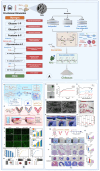
Tissue engineering applications and decellularized ECM procurement. (A) The secretive and ECM proteins found in the ECM promote cell division and proliferation. Alternative resources for tissue grafting are provided by the tissue structure and circulatory network that remain after the whole organ is decellularized. The dECM has many applications in different tissue engineering applications produced with different techniques, such as 3D-Bioprinting and electrospinning (created with Biorender.com). (B1B10) Schematic of experimental procedures. (B1B3) Isolation of fresh amniotic membrane (fAM) from fetal membrane. (B4B6) Preparation of decellularized and lyophilized amniotic membrane (DL-AM). (B7B12) Surgical procedures: endometrial injury in rats to induce IUA and DL-AM transplantation. (B13) Characterization of fAM vs. DL-AM by gross imaging, light microscopy, SEM, and H&E. (B14) Fibrosis evaluation post-surgery (Van Gieson staining at 3, 7, 14, and 28 days shows reduced fibrosis in the DL-AM group). (B15) Quantified fibrotic area confirms significant reduction vs. IUA group (* p < 0.05), but still higher than control (Copyright: © 2021 Chen et al. (Spandidos), licensed under CC BY-NC-ND 4.0) [103].
Figure 7
Figure 7
Structure and sources of cellulose from plants and bacteria. (A) Cellulose exhibits high biocompatibility, bioadhesiveness, biodegradability, and immunomodulatory properties, making it suitable for biomedical applications (created with Biorender.com). (B1,B2) Gel fraction (B1) and swelling ratio (B2) of CMC/gelatin hydrogels with varying compositions and radiation doses. B3–B4: In vitro enzymatic degradation of CMC/gelatin hydrogels (B3) and residual weight after 40 h in protease at 30 and 60 kGy (B4). (B5B7) Stress–strain curves (B5), compressive moduli (B6), and SEM cross-sectional images (B7) of hydrogels with varying compositions and irradiation doses. (B8B9) Cell viability and number of 3T3-Swiss fibroblasts on hydrogels (B8), and representative fluorescence images of remaining adhered cells (B9) (reproduced with permission, Elsevier) [141]. The significance levels are indicated as follows: *** for p < 0.001, ** for p < 0.01, * for p < 0.05, and ns for non-significant differences.
Figure 9
Figure 9
(A) Biosynthesis pathway of alginate, structure and ionotropic gelation mechanism of alginate, and applications in tissue engineering (created with Biorender.com). (B1) Preparation schematic of PVP-Alg and PVP-Alg-Ca films. (B2B6) Film morphology and SEM: porous vs. smooth surfaces and cell adhesion. (B7B9) Dissolution behavior and surface changes over time. (B10B15) Surgery site views and gene expression (TGF-β, TIMP-2); p < 0.05. (B16B24) Histology of injured sites (H&E, Masson, Sirius Red) showing tissue structure. (B25B28) Adhesion formation and histological origin in treatment groups (Copyright: © 2023 (Copyright: © 2023 Mao et al. (Frontiers), licensed under CC BY 4.0 et al. (MDPI—Materials), licensed under CC BY 4.0)) [191].
Figure 11
Figure 11
Elimination routes of monomers and biopolymers in the human body [257,258,259,260,261,262,263,264] (created with Biorender.com).
Figure 1
Figure 1
Collagen in anti-adhesion barriers and endometrial regeneration. (A) Structural representation of collagen. (B) Sources of collagen from animal and marine origins and applications of animal- and marine-derived collagens in anti-adhesion barrier system in uterine reconstruction (created with Biorender.com). (C1C6) Morphological and histological characterization of the collagen scaffold (CS) before and after UC-MSC seeding, showing porous structure, effective cell attachment, and proliferation. (C7C9) UC-MSCs enhanced HESC proliferation and reduced apoptosis in co-culture. (C10C12) ELISA confirmed elevated secretion of pro-regenerative factors (VEGF-A, TGF-β1, PDGF-BB). (D1D7) Gross views of uteri and embryos indicate restored fertility in the CS/UC-MSC group, with higher implantation rates compared to control groups (reproduced with permission, Elsevier). The significance levels are indicated as follows: ** for p < 0.01 and * for p < 0.05.
Figure 2
Figure 2
(A) Collagen triple-helix acid and base hydrolysis to break down bonding to form gelatin. (B) Triple helices supported by intermolecular hydrogen bonds cause a gelatin solution to gel when cooled; heating the resultant gelatin gel causes the opposite effect. (C) Application of gelatin as a biomaterial (created with Biorender.com). (D1D5) Macroscopic and histological assessment of abdominal adhesions 14 days post-surgery. (D1) Representative images of sham, adhesion, and gelatin groups. (D2) Adhesion scores (0–4 scale) show reduced adhesions in the gelatin group. (D3 a-f) Adhesion tissues from the sham, adhesion, and gelatin groups were stained with H&E. Newly formed blood vessels (arrowheads) and multinucleated giant cells (arrows) are visible. Scale bars: (A) a = 500 μm; b, c, e = 100 μm; d, f = 20 μm. (D4) Inflammatory scores (0–3 scale) and (D5) neovessel counts confirm lower inflammation and vascularization in the gelatin group (reproduced with permission, Elsevier) [48]. (E1E13) Characterization and evaluation of PU GF anti-adhesion film. (E1E4) Surface topography and structure of PU GF by roughness profiling and SEM imaging. (E5E6) Rat cecum abrasion model showing injury sites and sutured adhesion setup. (E7E9) PU GF exhibits superior tensile strength, elasticity, and enhanced fibroblast proliferation over time. (E10E13) H&E-stained sections reveal reduced inflammation and fibrotic response with Flat GF and PU GF compared to control and conventional films (Copyright: © 2025 Ozamoto et al. (PLoS One), licensed under CC BY 4.0) [49]. The significance levels are indicated as follows: ** for p < 0.01, * for p < 0.05.
Figure 3
Figure 3
(A) Fibrinogen assembly structure (created with Biorender.com). (B) TachoSil® (Takeda Pharmaceutical Company, Ltd., Osaka, Japan) structure. (C) Postoperative and histological evaluation of intrauterine adhesions. (C1C4) Laparoscopic views showing varying adhesion severity, from dense uterine adhesions to adhesion-free healing. (C5) Laparotomic view during hysterectomy and peritoneal biopsy in 49 rats/group to assess early effects of TachoSil®. (C6C7) Histology shows reduced uterine fibrosis and inflammation with TachoSil®, but no peritoneal improvement due to bipolar coagulation injury (UH uterine horn, UC uterine corpus, N necrosis, arrows inflammation) (adapted with permission, Springer) [67]. (D1D30) Evaluation of PRF structure and its effects on endometrial repair in a rat IUA model. (D1D9) PRF showed a distinct fibrin network and platelet-rich zones by histology, SEM, and TEM. (D10D30) PRF transplantation promoted epithelial regeneration, increased gland numbers, and reduced fibrosis compared to the IUA group, with outcomes approaching the sham group by day 14 (Copyright: © 2023 Mao et al. (Frontiers), licensed under CC BY 4.0) [68]. The significance levels are indicated as follows: *** for p < 0.001, ** for p < 0.01 and * for p < 0.05.
Figure 4
Figure 4
(A) Silk fibroin structure, synthesis, fabrication, and application as a natural biopolymer (created with Biorender.com). (B1B5) Physical properties of HMW and LMW SF hydrogels, including gelation (B1), light transmittance (B2), SEM (B3), FTIR (B4), and XRD patterns (B5). B6–B8: Biodegradation analysis of SF hydrogels via in vitro (B6, B7) and in vivo (B8) studies, showing faster degradation in LMW SF (* p < 0.05, *** p < 0.0001). (B9B11) Anti-adhesion effects of SF hydrogels in rats at day 7, with lower adhesion scores in LMW SF (B9), supported by gross (B10) and histological images (B11) (C, A, F, and asterisks indicate cecum, abdominal wall, fibrous tissue, and remaining SF hydrogels, respectively.) (reproduced with permission, ACS) [89].
Figure 6
Figure 6
(A) Illustration of the hyaluronan (HA) synthesis pathway. Glucose enters the cell via an ABC transporter and is converted to glucose-6-phosphate (1), followed by fructose-6-phosphate (2). Through a series of enzymatic reactions, these intermediates lead to the formation of UDP-glucuronic acid and UDP-N-acetylglucosamine (UDP-GlcNAc) (3). These two precursors are used by the enzyme hyaluronan synthase (4) to polymerize hyaluronan, which is then transported out of the cell. The figure also depicts different sources of HA (bacterial and animal) and its various biomedical applications, including drug carriers, electrospun membranes, and hydrogels in 3D biofabrication (created with Biorender.com). (B1) sHAChiF hydrogel showed rapid gelation, strong viscoelasticity, and effective sprayability with uniform coverage using a custom dual-spray system. (B2) sHAChiF showed low inflammation (via luminol assay) and sustained in vivo retention of both sHA and chitosan for up to 14 days, confirmed by IVIS imaging and fluorescence quantification. (B3) sHAChiF reduced macrophage aggregation and inflammation at injury sites, with strong localization of sHA and chitosan. Flow cytometry showed preferential sHA uptake by small peritoneal macrophages, supporting targeted anti-inflammatory effects (Copyright: © 2024 Song et al. (Springer Nature), licensed under CC BY-NC-ND 4.0) [120]. (C1) Hyalobarrier® (Gel A) (Anika Therapeutics, Inc., Bedford, MA, USA) showed significantly higher viscosity across shear rates compared to HyaRegen® (BioRegen Biomedical (Changzhou) Co., Ltd., Changzhou, China) and MateRegen® (BioRegen Biomedical (Changzhou) Co., Ltd., Changzhou, China), indicating superior mechanical stability. (C2) Hyalobarrier® exhibited the highest peel strength among the tested HA gels, indicating stronger tissue adhesion than HyaRegen® and MateRegen®. (C3) Peel strip images showed less residue and tearing with Hyalobarrier®, confirming its stronger and more cohesive adhesion compared to HyaRegen® and MateRegen® after repeated tests. (C4) HyaRegen® required the highest average extrusion force, while MetaRegen® required the lowest, indicating easier application for MetaRegen® but greater viscosity for HyaRegen® [121].
Figure 8
Figure 8
(A) Synthesis of the pathways of chitin in crustacean biowastes and extracted chitosan (created with Biorender.com). (B1,B2) Design of PEGMA/L-serine/PRP hydrogel and its anti-adhesion and PRP-release function. (B3B5) Cell viability, live/dead staining, and CCK-8 assay confirm good biocompatibility. (B6B8) Hydrogel shows controlled degradation, sustained VEGF/PDGF release, and in vivo degradation confirmed over 30 days (reproduced with permission, Elsevier) [150]. (C1C7) tHA-tChi hydrogel shows sol–gel transition, injectability, self-healing, and stable rheology. (C8C14) In vivo, it improves endometrial thickness, gland number, and reduces fibrosis (reproduced with permission, Elsevier) [151]. The significance levels are indicated as follows: **** p < 0.0001, *** for p < 0.001, ** for p < 0.01, * for p < 0.05, and ns for non-significant differences.
Figure 10
Figure 10
Structure and synthesis pathways of (A) PCL, (B) PEG, and (C) PLGA synthetic biopolymers commonly used in tissue engineering, along with their degradation mechanisms. Key factors influencing the rate and process of degradation, such as molecular weight. (D) represents the techniques and biomedical applications of PCL-PEG, and PLGA in tissue engineering (created with Biorender.com).
Figure 12
Figure 12
Schematic of conventional scaffold fabrication techniques: (A) gas foaming, (B) freeze drying, (C) porogen leaching, (D) phase separation, (E) melt molding (created with Biorender.com).

Similar articles

Cited by

References

    1. Weibel M.A., Majno G. Peritoneal adhesions and their relation to abdominal surgery: A postmortem study. Am. J. Surg. 1973;126:345–353. doi: 10.1016/S0002-9610(73)80123-0. - DOI - PubMed
    1. Lauder C.I., Garcea G., Strickland A., Maddern G.J. Abdominal adhesion prevention: Still a sticky subject. Dig. Surg. 2010;27:347–358. doi: 10.1159/000314805. - DOI - PubMed
    1. Schaefer S.D., Alkatout I., Dornhoefer N., Herrmann J., Klapdor R., Meinhold-Heerlein I., Meszaros J., Mustea A., Oppelt P., Wallwiener M., et al. Prevention of peritoneal adhesions after gynecological surgery: A systematic review. Arch. Gynecol. Obstet. 2024;310:655–672. doi: 10.1007/s00404-024-07584-1. - DOI - PMC - PubMed
    1. Wei Z., Hu Y., He X., Ling W., Yao J., Li Z., Wang Q., Li L. Biomaterializing the advances in uterine tissue engineering. iScience. 2022;25:105657. doi: 10.1016/j.isci.2022.105657. - DOI - PMC - PubMed
    1. Xu J., Fang H., Zheng S., Li L., Jiao Z., Wang H., Nie Y., Liu T., Song K. A biological functional hybrid scaffold based on decellularized extracellular matrix/gelatin/chitosan with high biocompatibility and antibacterial activity for skin tissue engineering. Int. J. Biol. Macromol. 2021;187:840–849. doi: 10.1016/j.ijbiomac.2021.07.162. - DOI - PubMed

LinkOut - more resources