Review
. 2025 Jun 9;11(6):442.
doi: 10.3390/gels11060442.

Recent Progress of Biomaterial-Based Hydrogels for Wearable and Implantable Bioelectronics

Affiliations
Review

Recent Progress of Biomaterial-Based Hydrogels for Wearable and Implantable Bioelectronics

Baojin Chen et al. Gels. .

Abstract

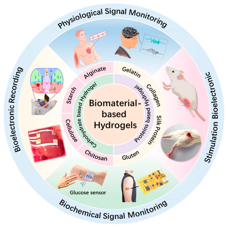
Bioelectronics for wearable and implantable biomedical devices has attracted significant attention due to its potential for continuous health monitoring, early disease diagnosis, and real-time therapeutic interventions. Among the various materials explored for bioelectronic applications, hydrogels derived from natural biopolymers have emerged as highly promising candidates, owing to their inherent biocompatibility, mechanical compliance akin to biological tissues, and tunable structural properties. This review provides a comprehensive overview of recent advancements in the design and application of protein-based hydrogels, including gelatin, collagen, silk fibroin, and gluten, as well as carbohydrate-based hydrogels such as chitosan, cellulose, alginate, and starch. Particular emphasis is placed on elucidating their intrinsic material characteristics, modification strategies to improve electrical and mechanical performance, and their applicability for bioelectronic interfaces. The review further explores their diverse applications in physiological and biochemical signal sensing, bioelectric signal recording, and electrical stimulation. Finally, current challenges and future perspectives are discussed to guide the ongoing innovation of hydrogel-based systems for next-generation bioelectronic technologies.

Keywords: bioelectronics; biomaterials; hydrogel; implantable; wearable.


Conflict of interest statement

The authors declare no conflicts of interest.

Figures

Figure 1
Figure 1
Recent progress of biomaterial-based hydrogels for wearable and implantable bioelectronics. Physiological Signal Monitoring: reproduced with permission [31]. Copyright 2023, Elsevier. Reproduced with permission [32]. Copyright 2023, Wiley. Stimulation Bioelectronic: reproduced with permission [33]. Copyright 2023, Springer Nature. Reproduced with permission [34]. Copyright 2025, Elsevier. Biochemical Signal Monitoring: reproduced with permission [35]. Copyright 2023, Cell Press. Reproduced with permission [36]. Copyright 2023, Elsevier. Bioelectronic Recording: reproduced with permission [37]. Copyright 2023, Springer Nature. Reproduced with permission [15]. Copyright 2024, Springer Nature.
Figure 2
Figure 2
Fabrication procedures and functional demonstrations of gelatin-based hydrogels. (a) Schematic illustration of the fabrication process for the multilayered chromotropic OE-skin [65]. (b) Synthetic route of PGAOH. (c) Functional demonstration matrix showcasing the multi-responsive properties of PGAOH [63]. (d) Schematic representation of the preparation process of PPG hydrogel. (e) Photographs of PPG hydrogel attached to human joints [66]. All pictures have been adopted with permission.
Figure 3
Figure 3
Structural design and functional demonstration of collagen-based hydrogels. (a) Schematic illustration of the fabrication process for CDPAP hydrogels [23]. (b) Photographs showing towel-inspired mechanical manipulation, including twisting and squeezing deformations. (c) Hierarchical multiscale crosslinking network in natural collagen fibrils. (d) Bioinspired processing strategy for collagen hydrogels [72]. All pictures have been adopted with permission.
Figure 4
Figure 4
Fabrication principles and morphological characteristics of silk protein-based hydrogels. (a) Schematic illustration of Ca2+-induced structural transition in silk proteins. (b) Micro-/nanopatterning technique employed in the fabrication of silk-based bioelectronic devices. (c) Optical image demonstrating the direct integration of silk bioelectronics. (d) Cross-sectional SEM image between red-dyed silk and PDMS [75]. (e) Schematic illustration of the synthesis route of the on-skin-formed silk protein (OSF-SP) bioelectrode and its secondary structure transition in the gelation process. (f) Photographs of a preformed film and an in situ-formed film covering the surface of a ball [55]. All pictures have been adopted with permission.
Figure 5
Figure 5
Fabrication strategy and multifunctional performance of glutenin-based hydrogels. (a) Schematic illustration of the preparation procedure of the gluten-based e-skin. (b) Photographs of stretched gluten hydrogel. (c) Different shapes of gluten hydrogel photographs [79]. (d) Schematic illustration of the fabrication process of conductive i-Gluten hydrogels. (e) Optical and SEM images displaying the morphology of i-Gluten. (f) Photographs demonstrating the excellent stretchability of i-Gluten. (g) Photographs showing the adhesive capability of i-Gluten on various substrates. (h) Photographs highlighting the self-recovery performance of i-Gluten after mechanical damage [80]. All pictures have been adopted with permission.
Figure 6
Figure 6
Fabrication strategies and structural characterization of chitosan-based hydrogels. (a) Schematic illustration of the fabrication process for PHA/x-CS hydrogels. (b) Representative tensile stress–strain curves comparing PHA/CS and PHA/x-CS hydrogels. (c) Schematic of the photomask-assisted patterning approach for PHA/x-CS hydrogels. (d) The mechanical deformation of the patterned hydrogel under stretching. (e) Optical transmittance spectrum of the PHA/x-CS hydrogel [42]. (f) Schematic of in situ 3D printing of LM-hydrogel hybrids. (g) Elastic modulus comparison between GSP hydrogel and LM-hydrogel hybrid. (h) Twist deformation comparison of LM–hydrogel and metal–hydrogel composites [34]. All pictures have been adopted with permission.
Figure 7
Figure 7
Preparation strategies and structural characterization of cellulose-based hydrogels. (a) Schematic of hierarchical microstructure in cellulose/BT hydrogels. (b) Photograph of the synthesized ion–CB hydrogel. (c) Optical image demonstrating the scalable fabrication of ion–CB hydrogels [99]. (d) Schematic of fabrication and dynamic network structure of PAM/PBA-IL/CNF hydrogels. (e) Tensile deformation of PAM/PBA-IL/CNF hydrogels under stress. (f) Design schematic of PAA-CNF-IL-H2O ionogels. (g) Visual of tensile performance of PAA-CNF-IL-H2O ionogels. (h) Molecular schematic of noncovalent bonding in PAA-CNF-IL-H2O ionogels [101]. All pictures have been adopted with permission.
Figure 8
Figure 8
Structural mechanisms and enhanced mechanical performance of alginate-based hydrogels. (a) Schematic illustration of ionic crosslinking in Alg hydrogels. (b) Covalent crosslinking architecture in PAAm hydrogels formed via free-radical polymerization. (c) Hybrid Alg/PAAm network with ionic and covalent crosslinks. (d) Mechanical performance of the hybrid hydrogel [43]. (e) Schematic of dual crosslinking in Alg/PVA hydrogels. (f) Reinforcement strategy using ionic crosslinking and salting-out in Alg/PVA hydrogels. (g) Visual comparison of Alg/PVA hydrogels in different treatment states [106]. All pictures have been adopted with permission.
Figure 9
Figure 9
Preparation strategies and characterization of starch-based hydrogels. (a) Schematic of AAM hydrogel fabrication process. (b) Photographs of AAM hydrogels before and after thermal treatment. (c) Optical transmittance spectra of AAM hydrogels [114]. (d) Formation mechanism of CS-SA-Ca2+ hydrogel via ionic crosslinking. (e) Tensile deformation behavior of CS-SA-Ca2+ hydrogel [108]. All pictures have been adopted with permission.
Figure 10
Figure 10
Applications of biomaterial-based hydrogels in physiological signal monitoring. (a) Schematic of PAC2V3 hydrogel electrodes for ECG monitoring. (b) Diagram of the ECG monitoring mechanism. (c) ECG signals from PAC2V3 electrodes under three physiological states [31]. (d) Structural layout of the EEG monitoring system employing hydrogel-based components. (e) Comparative analysis of motion artifacts between P-P-PDA nanoparticle electrodes and conventional wet Ag/AgCl electrodes [32]. (f) Setup for facial EMG recording with hydrogel electrodes. (g) EMG signals for different facial expressions. (h) Comparison of SNR of Ag/AgCl and hydrogel electrodes under different conditions [141]. All pictures have been adopted with permission.
Figure 11
Figure 11
Hydrogel-based biochemical sensors and their physiological monitoring capabilities. (a) Schematic of a wearable device for UA monitoring. (b) Photographs of the UA sensor patch and its on-skin application [35]. (c) Diagram of a self-healing, glucose-adaptive hydrogel triboelectric sensor for sweat analysis [36]. (d) System connectivity of the CPPH sweat sensor with wireless components. (e) Real-time wireless sensing performance during running. (f) Ion detection profiles (Na+, K+, Ca2+) over 30 min of exercise [160]. All pictures have been adopted with permission.
Figure 12
Figure 12
Biomaterial-based hydrogel electrodes for implantable electrophysiological recording. (a) Schematic of an in vivo material performance testing setup. (b) Images showing ultraconformal adhesion of the SMCA sensor on an ex vivo bovine cortex [15]. (c) Diagram of a SAFIE system on a rat heart. (d) Photographs of SAFIE deployed on cardiac tissue. (e) Real-time ECG recordings from four channels showing ventricular activity 4 weeks after implantation [37]. (f) Photograph of a viscoelastic array placed on rat cortical dura. (g) Schematic and data showing flexible array deployment and auditory cortex responses to sound stimulation [50]. All pictures have been adopted with permission.
Figure 13
Figure 13
Schematic illustration and functional characterization of implantable electrical stimulation systems utilizing biomaterial hydrogel electrodes. (a) Photograph of neural stimulation using IT-IC hydrogel interfaces. (b) Correlation between leg vibration and stimulation frequency with motion snapshots. (c) Photographs showing ankle angle changes under different voltages [33]. (d) Schematic illustrations and an image of the sciatic nerve stimulation setup. (e) Physical attachment of a stimulation electrode on a sciatic nerve without the e-bioadhesive interface. (f) Robust integration of a stimulation electrode on a sciatic nerve with the e-bioadhesive interface. (g) Images of the ankle joint movement in response to electrical stimulation via the e-bioadhesive interface [172]. (h) Schematic of TA muscle stimulation using LM–hydrogel hybrid electrodes in rats. (i) Comparative images of ankle joint kinematics during TA muscle stimulation, employing LM–hydrogel electrodes versus pure GSP hydrogel controls. (j) Force output measurements under different TA muscle stimulation conditions [34]. All pictures have been adopted with permission.

Similar articles

Cited by

References

    1. Koo J., MacEwan M.R., Kang S.K., Won S.M., Stephen M., Gamble P., Xie Z., Yan Y., Chen Y.Y., Shin J., et al. Wireless bioresorbable electronic system enables sustained nonpharmacological neuroregenerative therapy. Nat. Med. 2018;24:1830–1836. doi: 10.1038/s41591-018-0196-2. - DOI - PubMed
    1. Tringides C.M., Mooney D.J. Materials for Implantable Surface Electrode Arrays: Current Status and Future Directions. Adv. Mater. 2022;34:e2107207. doi: 10.1002/adma.202107207. - DOI - PubMed
    1. Khan W.U., Shen Z., Mugo S.M., Wang H., Zhang Q. Implantable hydrogels as pioneering materials for next-generation brain-computer interfaces. Chem. Soc. Rev. 2025;54:2832–2880. doi: 10.1039/D4CS01074D. - DOI - PubMed
    1. Chung H.U., Kim B.H., Lee J.Y., Lee J., Xie Z., Ibler E.M., Lee K., Banks A., Jeong J.Y., Kim J., et al. Binodal, wireless epidermal electronic systems with in-sensor analytics for neonatal intensive care. Science. 2019;363:eaau0780. doi: 10.1126/science.aau0780. - DOI - PMC - PubMed
    1. Lu Y., Yang G., Wang S., Zhang Y., Jian Y., He L., Yu T., Luo H., Kong D., Xianyu Y., et al. Stretchable graphene–hydrogel interfaces for wearable and implantable bioelectronics. Nat. Electron. 2023;7:51. doi: 10.1038/s41928-023-01091-y. - DOI

LinkOut - more resources