Fracture Resistance of CAD/CAM-Fabricated Zirconia and Lithium Disilicate Crowns with Different Margin Designs: Implications for Digital Dentistry
- PMID: 40558892
- PMCID: PMC12193716
- DOI: 10.3390/jfb16060205
Fracture Resistance of CAD/CAM-Fabricated Zirconia and Lithium Disilicate Crowns with Different Margin Designs: Implications for Digital Dentistry
Abstract
Objective: This in vitro study aimed to evaluate the influence of cervical margin design-tangential versus chamfer-on the fracture resistance of monolithic crowns fabricated from lithium disilicate and zirconia ceramics.
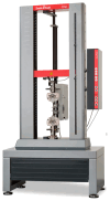
Materials and methods: Forty extracted human molars were randomly assigned to two preparation types: chamfer and tangential. Each group was restored with CAD/CAM-fabricated crowns made from either zirconia (IPS e.max® ZirCAD Prime) or lithium disilicate (IPS e.max® CAD), resulting in four subgroups (n = 10). Standardized adhesive cementation protocols were applied. After 24 h storage in distilled water, the specimens underwent static load-to-failure testing using a ZwickRoell ProLine Z005 universal testing machine.
Results: Zirconia crowns with chamfer margins exhibited the highest mean fracture resistance (2658 N), while lithium disilicate crowns with tangential margins showed the lowest (1862 N). Chamfer preparation significantly increased the fracture resistance of lithium disilicate crowns (p < 0.01), whereas margin design had no significant effect on zirconia. All restorations exceeded physiological masticatory forces, confirming their clinical viability.
Conclusions: Cervical margin design significantly affected the fracture performance of lithium disilicate crowns but not zirconia. Chamfer preparations are recommended when using lithium disilicate to optimize mechanical strength. These findings underscore the importance of preparation geometry in guiding material selection for CAD/CAM ceramic restorations.
Keywords: CAD/CAM; adhesive cementation; cervical margin; chamfer; fracture resistance; lithium disilicate; tangential; zirconia crowns.
Conflict of interest statement
The authors declare no conflicts of interest.
Figures


Similar articles
-
Adhesive Performance of Zirconia and Lithium Disilicate Maryland Cantilever Restorations on Prepared and Non-Prepared Abutment Teeth: An In Vitro Comparative Study.Biomimetics (Basel). 2025 Jun 21;10(7):413. doi: 10.3390/biomimetics10070413. Biomimetics (Basel). 2025. PMID: 40710226 Free PMC article.
-
Fracture resistance of chairside cad/cam advanced lithium disilicate maxillary canine veneers with different incisal edge designs.Saudi Dent J. 2025 Jul 1;37(4-6):27. doi: 10.1007/s44445-025-00029-8. Saudi Dent J. 2025. PMID: 40593259 Free PMC article.
-
Effect of finish line designs on the dimensional accuracy of monolithic zirconia crowns fabricated with a material jetting technique.J Prosthet Dent. 2025 Jul;134(1):176.e1-176.e8. doi: 10.1016/j.prosdent.2025.02.055. Epub 2025 Mar 17. J Prosthet Dent. 2025. PMID: 40102167
-
Metal-free materials for fixed prosthodontic restorations.Cochrane Database Syst Rev. 2017 Dec 20;12(12):CD009606. doi: 10.1002/14651858.CD009606.pub2. Cochrane Database Syst Rev. 2017. PMID: 29261853 Free PMC article.
-
Analysis of Different Lithium Disilicate Ceramics According to Their Composition and Processing Technique-A Systematic Review and Meta-Analysis.Materials (Basel). 2025 Jun 9;18(12):2709. doi: 10.3390/ma18122709. Materials (Basel). 2025. PMID: 40572842 Free PMC article. Review.
Cited by
-
Adhesive Performance of Zirconia and Lithium Disilicate Maryland Cantilever Restorations on Prepared and Non-Prepared Abutment Teeth: An In Vitro Comparative Study.Biomimetics (Basel). 2025 Jun 21;10(7):413. doi: 10.3390/biomimetics10070413. Biomimetics (Basel). 2025. PMID: 40710226 Free PMC article.
References
-
- Hategan S.I., Belea A.L., Tanase A.D., Gavrilovici A.M., Petrescu E.L., Marsavina L., Sinescu C., Negrutiu M.L., Manole M.C. The Evaluation of the Prosthetic Preparations Finish Lines for Provisional Crowns with the Impact on Periodontal Health. Rom. J. Oral Rehabil. 2024;16:453–459. doi: 10.62610/RJOR.2024.3.16.47. - DOI
LinkOut - more resources
Full Text Sources
Miscellaneous

