. 2025 May 30;11(6):423.
doi: 10.3390/jof11060423.

Histopathological Study of Host-Pathogen Interactions Between Cordyceps javanica PSUC002 and Hypothenemus hampei

Affiliations

Histopathological Study of Host-Pathogen Interactions Between Cordyceps javanica PSUC002 and Hypothenemus hampei

Sinlapachai Senarat et al. J Fungi (Basel). .

Abstract

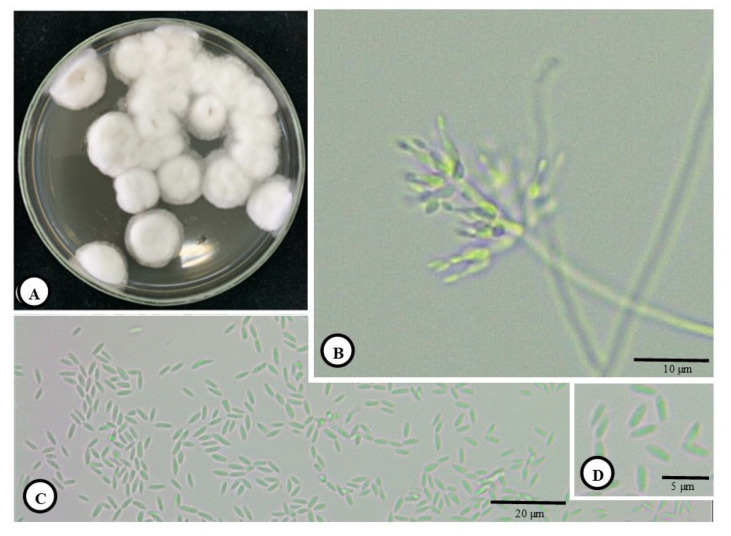
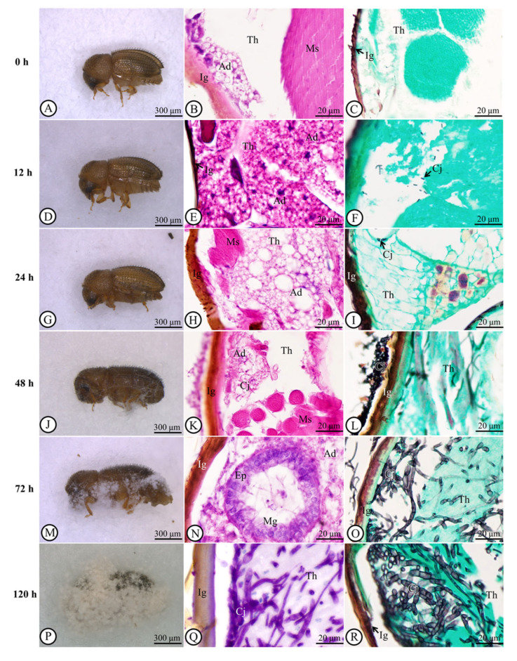
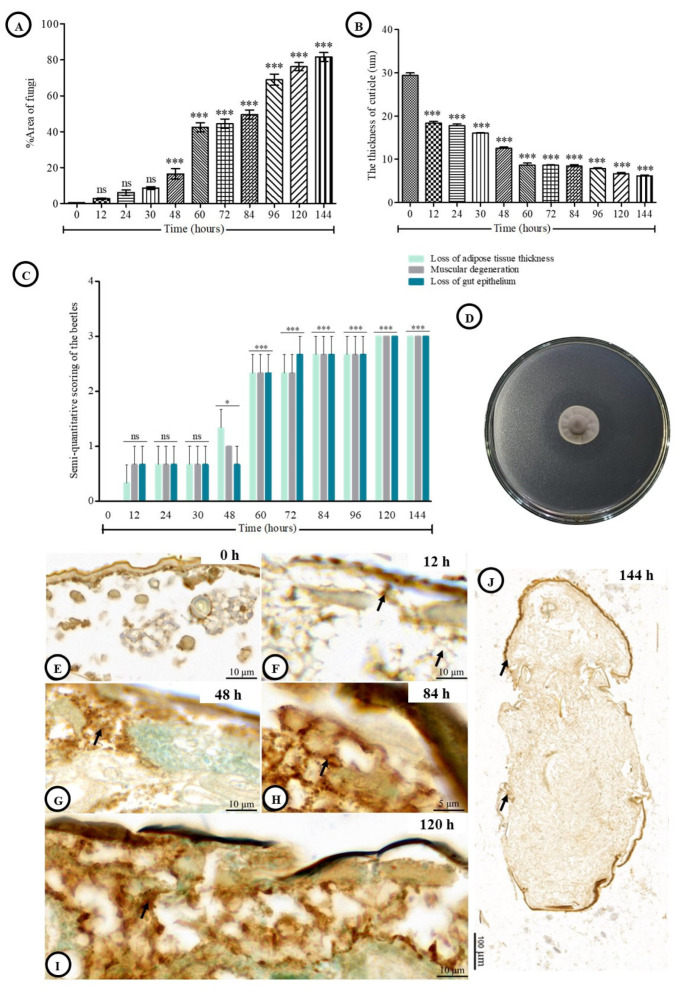
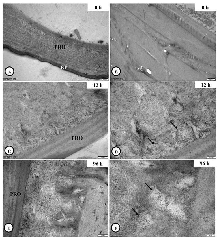
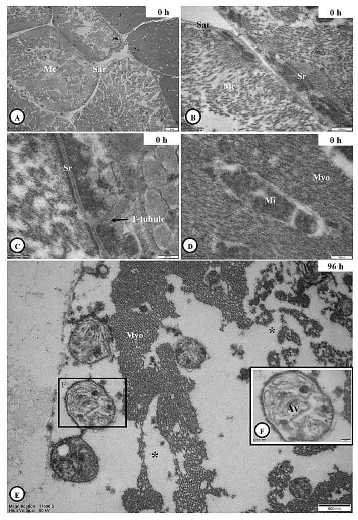
The use of entomopathogenic fungi (EPF), such as Cordyceps javanica, to reduce insect pest populations is gaining traction since it is an environmentally safe approach that can control many pests at different life stages. Here, we focus on the histopathology of the coffee berry borer, Hypothenemus hampei, infected by C. javanica. Morphological observation revealed that C. javanica conidia germinated within 12 h following inoculation according to light microscopic and ultrastructural levels. The fungus thoroughly penetrated the fat body and muscular tissue between 84 and 120 h post-inoculation. Transmission electron microscopy (TEM) confirmed the hyphal invasion of the cuticle at 12 h post-inoculation, with progressive tissue disruption and organelle degeneration, especially mitochondria and rough endoplasmic reticulum in adipocytes. All organelles were completely degenerated at 96 h post-inoculation. There was evidence of a connection between C. javanica activity and the coffee berry borer that might cause histopathological changes in the host defense against the pathogen, pointing to increased mortality and potential control of coffee berry borer in natural populations. Additionally, terminal deoxynucleotidyl transferase dUTP nick end labeling (TUNEL) confirmed that apoptotic cells were slightly increased in the adipose tissue and integument of the coffee berry borer. The ability of C. javanica to fatally infect the coffee berry borer suggests that it could be deployed as a biological control agent in the field.

Keywords: Cordyceps javanica; coffee berry borer; conidia infestation; insect integument.


Conflict of interest statement

The authors declare no conflict of interest.

Figures

Figure 1
Figure 1
Morphology of C. javanica, (A): Colony of cultured C. Javanica. (B): Phialides formation of C. Javanica and (C,D): separated conidia spores.
Figure 2
Figure 2
Histopathological development of C. javanica in a H. hampei host. (A): Adult H. Hampei. (B): Normal integument structure by H&E staining. (C): GMS stain performed on integument tissue. (D): At 12 h post-inoculation, hyphal invasion of the fat body underlying the cuticle stained with H&E (E) and GMS (F). (G): At 24 h post-inoculation, hyphal invasion of the fat body underlying the cuticle, stained with H&E (H) and GMS (I). (J): At 48 h post-inoculation, hyphal invasion of fat body underlying the cuticle, stained with H&E (K) and GMS (L). (M): At 72 h post-inoculation, hyphal invasion of fat body underlying the cuticle, stained with H&E (N) and GMS (O). (P): At 120 h post-inoculation, hyphal invasion of fat body covering the cuticle, stained with H&E (Q) and GMS (R). Staining methods: H&E = hematoxylin and eosin, GMS = Grocott’s methenamine silver stain. Abbreviations: Ad = adipose tissue, Cj = C. javanica, Ep = epithelium, h = hour, Ig = integument, Mg = midgut, Ms = muscle, Th = thorax.
Figure 3
Figure 3
The bar graph and Light microscope observation of apoptotic cells (arrows) in adult H. hampei specimens. (A): A comparison of percentage areas of fungal infection of the control (0 h) and at various times after inoculation with C. javanica. Values are presented as means ± SE. Statistically highly significant (*** p < 0.001). (B): The thickness of the cuticle of H. hampei after inoculation with C. javanica. Values are presented as means ± SE. Statistically highly significant (*** p < 0.001). (C): Histopathological alteration indexes (HAI) of adult H. hampei specimens inoculated with C. javanica. Data compare the loss of adipose tissue thickness, muscular degeneration and loss of gut epithelium. Values are presented as means ± SE. Statistically highly significant (*** p < 0.001). (D): The lipase production in mycelial agar discs. (EJ): The apoptotic cells (arrows) in adult H. hampei specimens inoculated with C. javanica at different post-inoculation times: 0 h (E), 12 h (F), 48 h (G), 84 h (H), 120 h (I) and 144 h (J). Statistical significance: * p < 0.05 and ns = non-significant.
Figure 4
Figure 4
Ultrastructural integuments of adult H. hampei specimens after exposure to C. javanica. (A,B): A clear observation of integument having the procuticle (PRO) and epicuticle (EP) at 0 h post-inoculation. (C,D): A few penetrations of hyphae with an amorphous and electron-lucent (arrows) of C. javanica throughout the procuticle (PRO) at 12 h post-inoculation. (E,F): The well penetrations of germination and hyphal structure of C. javanica at 96 h post-inoculation.
Figure 5
Figure 5
Ultrastructural adipocyte of adult H. hampei specimens after exposure with C. javanica. (A,B): The adipocyte at 0 h post-inoculation with the occurrence of a large central nucleus and the well-development of rough endoplasmic reticulum (RER) together with the ribosomes (arrows), mitochondria (Mi) and vacuoles (V). (CE): The adipocyte at 12 h post-inoculation with the similar previous post-inoculation was observed; however, the RER, mitochondria (Mi) and ribosomes (arrows) were degenerated in some areas of this cell (F). (G): The adipocyte without the organelles above (single asterisk) at 96 h post-inoculation, whereas its nucleus with the fragmented nuclear membrane (FNm) and euchromatic necrosis (double asterisks) were identified (H). Abbreviations: Eu = euchromatin, Hc = heterochromatin, PRO = procuticle.
Figure 6
Figure 6
Ultrastructural muscle cells of adult H. hampei specimens after exposure with C. javanica. (A): The muscle cell (Mc) surrounding the sarcolemma (Sar) at 0 h post-inoculation was described, whereas its sarcoplasmic reticulum (Sr) was corrected (B). (C,D): High magnification showed the transverse (T-tubules are linked with the sarcoplasmic reticulum (Sr) and the mitochondria (Mi)-associated myofibrils (Myo) in the muscle cells. (E,F): The appearance of necrosis of myofibrils (*) with several autophagic vacuoles (Av) was observed at 96 h post-inoculation.

Similar articles

References

    1. Avelino J., Ten Hoopen G.M., DeClerck F. Ecological mechanisms for pest and disease control in coffee and cacao agroecosystems of the neotropics. In: Rapidel B., DeClerck F., Le Coq J.F., Beer J., editors. Ecosystem Services from Agriculture and Agroforestry: Measurement and Payment. 4th ed. Earthscan Publications; London, UK: 2011. pp. 91–117.
    1. Harelimana A., Rukazambuga D., Hance T. Pests and diseases regulation in coffee agroecosystems by management systems and resistance in changing climate conditions: A review. J. Plant Dis. Prot. 2022;129:1041–1052. doi: 10.1007/s41348-022-00628-1. - DOI
    1. Lamichhane J.R., Arendse W., Dachbrodt-Saaydeh S., Kudsk P., Roman J.C., van Bijsterveldt-Gels J.E.M., Wick M., Messéan A. Challenges and opportunities for integrated pest management in Europe: A telling example of minor uses. Crop Prot. 2015;74:42–47. doi: 10.1016/j.cropro.2015.04.005. - DOI
    1. Groenen D. The effects of climate change on the pests and diseases of coffee crops in mesoamerica. J. Climatol. Weather Forecast. 2018;6:239. doi: 10.4172/2332-2594.1000239. - DOI
    1. Rice R.A. Coffee in the crosshairs of climate change: Agroforestry as abatis. Agroecol. Sustain. Food Syst. 2018;42:1058–1076. doi: 10.1080/21683565.2018.1476428. - DOI

LinkOut - more resources