

A New Classification of Inferior Alveolar Nerve Repositioning Procedures for Dental Implant Placement
- PMID: 40559170
- PMCID: PMC12192414
- DOI: 10.3390/dj13060267
A New Classification of Inferior Alveolar Nerve Repositioning Procedures for Dental Implant Placement
Abstract
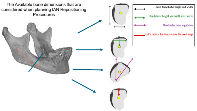
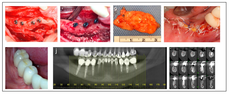
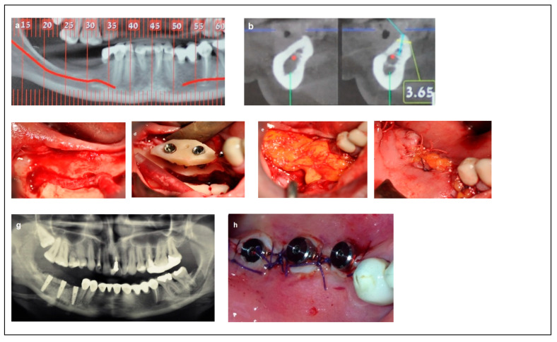
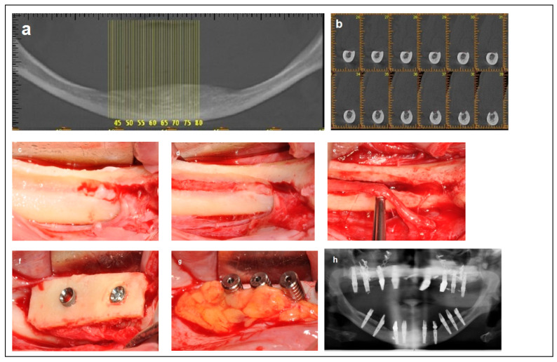
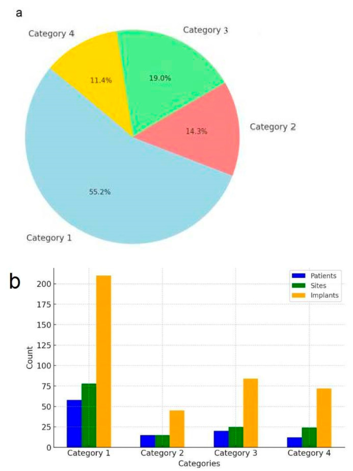
Background: Tooth loss significantly impacts the quality of life for adults. Inferior alveolar nerve (IAN) repositioning has garnered interest as a treatment for facilitating dental implant placement in the severely atrophic posterior mandible. However, there remains a need for standardization and classification of these techniques to improve outcomes. This study aims to propose a new clinical classification system for IAN repositioning procedures based on anatomical and procedural parameters. Methods: This study retrospectively analyzed preoperative radiographic records and surgical procedure documents over a 15-year period (2008-2023) for patients who underwent implant placement combined with IAN repositioning in the posterior atrophic mandible. Cases were classified into four categories according to bone availability, nerve location, and type of surgical intervention. Results: The study analyzed 142 edentulous posterior mandibles in 105 patients (77 women, 28 men; age range: 20-75). The cases were divided into four categories: Category 1 (58 patients, 78 sites), treated with one surgery; Category 2 (15 patients, 15 sites), treated in two stages; Category 3 (20 patients, 25 sites); and Category 4 (12 patients, 24 sites), with Categories 3 and 4 treated in a single surgery. Across all 132 sites, 411 dental implants were placed and restored with implant-supported fixed prostheses. Conclusions: This proposed classification provides a structured systematic framework for assessing and planning IAN repositioning procedures. It facilitates better diagnosis, treatment planning, and prediction of surgical stages in patients needing IAN repositioning for dental implant placement.
Keywords: atrophic mandible; bone grafts; dental implants; inferior alveolar nerve.
Conflict of interest statement
The author declares no conflicts of interest.
Figures
Similar articles
-
Interventions for replacing missing teeth: management of soft tissues for dental implants.Cochrane Database Syst Rev. 2012 Feb 15;2012(2):CD006697. doi: 10.1002/14651858.CD006697.pub2. Cochrane Database Syst Rev. 2012. PMID: 22336822 Free PMC article.
-
Effectiveness of sinus lift procedures for dental implant rehabilitation: a Cochrane systematic review.Eur J Oral Implantol. 2010 Spring;3(1):7-26. Eur J Oral Implantol. 2010. PMID: 20467595
-
Effect of maxillary sinus augmentation on the survival of endosseous dental implants. A systematic review.Ann Periodontol. 2003 Dec;8(1):328-43. doi: 10.1902/annals.2003.8.1.328. Ann Periodontol. 2003. PMID: 14971260
-
Surgical techniques used in the rehabilitation of partially edentulous patients with atrophic posterior mandibles: A systematic review and meta-analysis of randomized controlled clinical trials.J Craniomaxillofac Surg. 2017 Aug;45(8):1236-1245. doi: 10.1016/j.jcms.2017.04.011. Epub 2017 Apr 27. J Craniomaxillofac Surg. 2017. PMID: 28552200
-
Complications associated with inferior alveolar nerve repositioning for dental implant placement: a systematic review.Int J Oral Maxillofac Surg. 2014 Nov;43(11):1360-6. doi: 10.1016/j.ijom.2014.07.010. Epub 2014 Aug 12. Int J Oral Maxillofac Surg. 2014. PMID: 25128261
References
-
- Lorean A., Kablan F., Mazor Z., Mijiritsky E., Busse P., Barbu H.M., Levin L. Inferior alveolar nerve transposition and reposition for dental implant placement in edentulous or partially edentulous mandibles: A multicenter retrospective study. Int. J. Oral Maxillofac. Surg. 2013;42:656–659. doi: 10.1016/j.ijom.2013.01.020. - DOI - PubMed
-
- Barbu H.M., Levin L., Bucur M.B., Comaneanu R.M., Lorean A. A modified surgical technique for inferior alveolar nerve repositioning on severely atrophic mandibles: Case series of 11 consecutive surgical procedures. Chirurgia. 2014;109:111–116. - PubMed
LinkOut - more resources
Full Text Sources
