Review
. 2025 Jun 18;15(12):943.
doi: 10.3390/nano15120943.

Graphene-Based Plasmonic Antenna for Advancing Nano-Scale Sensors

Affiliations
Review

Graphene-Based Plasmonic Antenna for Advancing Nano-Scale Sensors

Waqas Ahmad et al. Nanomaterials (Basel). .

Abstract

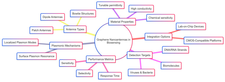
The integration of two-dimensional graphene with gold nanostructures has significantly advanced surface plasmon resonance (SPR)-based optical biosensors, due to graphene's exceptional optical, electronic, and surface properties. This review examines recent developments in graphene-based hybrid nanomaterials designed to enhance SPR sensor performance. The synergistic combination of graphene and other functional materials enables superior plasmonic sensitivity, improves biomolecular interaction, and enhances signal transduction. Key focus areas include the fundamental principle of graphene-enhanced SPR, the functional advantages of graphene hybrid platforms, and their recent applications in detecting biomolecules, disease biomarkers, and pathogens. Finally, current limitations and potential future perspectives are discussed, highlighting the transformative potential of these hybrid nanomaterials in next-generation optical biosensing.

Keywords: biomolecular interactions; hybrid nanomaterials; next-generation biosensing; plasmonic sensitivity.


Conflict of interest statement

The authors declare no conflicts of interest.

Figures

Figure 6
Figure 6
Design and optical response of a hybrid metal-graphene Fano-resonant metamaterial. (a) Schematic illustration of the proposed hybrid metamaterial structure. (bd) Simulated optical spectra showing transmission, reflection, and absorption for three configurations: (b) nanostructured gold film without graphene nano disks, (c) graphene nano disks without nanostructured gold, and (d) the complete hybrid metamaterial combining both components. (e,f) distributions of the local electric field in the z-direction at the resonance wavelength for (e) graphene nano disks (~10 μm) and (f) the hybrid structure (~10.05 μm). The field is normalized to the incident field amplitude E0 and plotted in the x-y plane located 5 nm above the graphene layer. An x-polarized plane wave is incident normally from the top side of the structure [93].
Figure 1
Figure 1
(top) Schematic illustration of localized surface plasmon (LSP) oscillations in a metal nanoparticle, where the electron cloud resonates with the incident electric field. (Bottom) Lossless dispersion curve for surface plasmon resonance (SPR). The red curve represents the dispersion of SPPs at a metal–dielectric interface, asymptotically approaching the light line. The flat blue line indicates the non-dispersive resonance frequency of LSPs. The light cone represents freely propagating photons in the dielectric [52].
Figure 2
Figure 2
Schematic showing the electromagnetic field distribution at a metal–dielectric interface in SPR sensors. The evanescent field decays exponentially into both materials, with a typical 200–500 nm penetration depth. This near-field interaction governs the sensitivity of SPR to local refractive index changes and results in measurable shifts in the resonance angle [62].
Figure 3
Figure 3
(a) Schematic illustration of the fabrication process for the graphene-PDMS (polydimethylsiloxane) flexible device. The process begins with a mold and electrode bonding, followed by graphene transfer and pouring of the PDMS solution. After curing and drying, the device is tailored into the final structure. (b) Electrical setup of the fabricated device showing the integrated graphene layer with gold electrodes embedded in PDMS, connected to a power supply and resistor for measurement [63].
Figure 4
Figure 4
Schematic of the two-slit plasmonic antenna milled in an Au film on top of a silica substrate, excited by a crossed femtosecond laser dual-beam [11].
Figure 5
Figure 5
(a) Schematic drawing of the LSPR sensing structure. (b) Hexagonal periodic dielectric-metal hybrid structure. (c) Structural side view. (d) The corresponding simulated transmission spectra in water with D = 300 nm, P = 450 nm, h1 = 120 nm, h2 = 80 nm, and Ng = 20, at normal incidence under TM polarization. The inset of the figure provides electric-field profiles corresponding to the dips [83].
Figure 7
Figure 7
Design, radiation patterns, and performance analysis of graphene-based plasmonic nanoantenna arrays. (A) Schematic views of a graphene square patch nanoantenna: (a) top view showing patch dimensions and feed width, (b) 3D perspective view, and (c) side view indicating material layering with SiO2 substrate and nanostrip line feed. (B) Radiation patterns of graphene nanoantenna arrays for different configurations: (a) 2 × 2, (b) 3 × 3, and (c) 4 × 4 arrays, showing vertically and horizontally polarized field distributions. (C) Comparative analysis of directivity (a) and gain (b) for square-shaped vs. L-shaped graphene nanoantenna arrays across increasing array dimensions [110].
Figure 8
Figure 8
Illustrates a graphene-based hyperbolic metamaterial (HMM) biosensor integrated with a gold grating structure for mid-infrared plasmonic sensing. (a) Design overview showing the Au grating coupled with a graphene/Al2O3 multilayer HMM stack under TM-polarized incident light. (b) Three-dimensional model of the unit cell with alternating graphene and Al2O3 bilayers (N = 11). (c) Top and side views highlighting the geometry of the grating and HMM layers. (d) Effective permittivity analysis showing real and imaginary components, indicating a hyperbolic dispersion regime beyond 3.58 μm [129].
Figure 9
Figure 9
(A) Schematic of different plasmonic nanoantenna geometries commonly used in SPR-based biosensing: bowtie, circular, and rectangular antenna pairs positioned on a dielectric substrate. Dimensions for each geometry are annotated, showing width, length, and interparticle gap. (B) Conceptual illustration of localized surface plasmon resonance (LSPR) excitation and field distribution on a graphene surface. The electric (E-field) and magnetic (H-field) components of the surface plasmon mode are shown, along with the propagation direction λsp and (ns) near-field intensity decay [140].
Figure 10
Figure 10
Schematic of a graphene-enhanced biosensor for cardiac biomarker detection. (A) Functionalize a gold screen-printed electrode (SPE) using electrophoretic deposition of graphene oxide/polyethylenimine (GO/PEI), followed by EDC/NHS activation. (B) Site-specific attachment of aptamers for BNP-32 and cTnI detection through analyte-induced changes in the sensor response. (I) Electrophoretic deposition of GO/PEI onto SPE. (II) Propargyl functionalization via EDC/NHS chemistry (III) click conjugation of azide-modified aptamers for BNP-32 or cTnI (IV) PEG modification for antifouling surface treatment [144].
Figure 11
Figure 11
Electrochemical detection of miRNA-21 using various modified electrode configurations. The DPV curves demonstrate signal enhancement from bare DCE through successive functionalization, with the final configuration miRNA-21/Apt/PEDOT/Pep/AuNP/GCE showing the highest current response, indicating superior sensitivity. This graphene-integrated platform enables efficient and label-free detection of microRNA biomarkers relevant to cancer diagnostics [188].
Scheme 1
Scheme 1
Advantages of graphene-based plasmonic nanoantennas in biosensing applications.

Similar articles

References

    1. Sangwan A., Jornet J.M. Beamforming optical antenna arrays for nano-bio sensing and actuation applications. Nano Commun. Netw. 2021;29:100363. doi: 10.1016/j.nancom.2021.100363. - DOI
    1. Gopalan K.K., Paulillo B., Mackenzie D.M., Rodrigo D., Bareza N., Whelan P.R., Shivayogimath A., Pruneri V. Scalable and tunable periodic graphene nanohole arrays for mid-infrared plasmonics. Nano Lett. 2018;18:5913–5918. doi: 10.1021/acs.nanolett.8b02613. - DOI - PubMed
    1. Huang Y.H., Ho H.P., Wu S.Y., Kong S.K. Detecting phase shifts in surface plasmon resonance: A review. Adv. Opt. Technol. 2012;2012:471957. doi: 10.1155/2012/471957. - DOI
    1. Sirenko Y.K., Strom S. Modern Theory of Gratings. Resonant Scattering: Analysis Techniques and Phenomena. Springer; Berlin/Heidelberg, Germany: 2010.
    1. Nguyen H.H., Park J., Kang S., Kim M. Surface Plasmon Resonance: A Versatile Technique for Biosensor Applications. Sensors. 2015;15:10481–10510. doi: 10.3390/s150510481. - DOI - PMC - PubMed

LinkOut - more resources