. 2025 Jun 1;8(3):56.
doi: 10.3390/mps8030056.

Towards Automated Testing of Kynurenine for Point-of-Care Metabolomics

Affiliations

Towards Automated Testing of Kynurenine for Point-of-Care Metabolomics

Dipanjan Bhattacharyya et al. Methods Protoc. .

Abstract

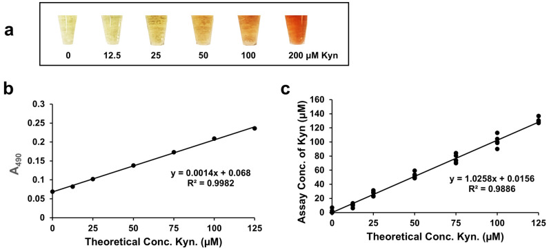
Our objective was to develop a simple, low-cost colorimetric assay to detect kynurenine (L-Kyn) in human biofluids, that would be compatible with a point-of-care (POC) system being developed in our lab. Elevated L-Kyn is associated with many pathological conditions. However, current detection methods are expensive, time-consuming, and unsuitable for resource-limited settings. Existing colorimetric L-Kyn assays lack specificity, require unusual reagents, or lack sensitivity, hindering their practical application. Here we report a two-step diazotization-based colorimetric assay that produces a red chromophore upon reaction with L-Kyn. To reduce background interference, we used dilution and anion exchange chromatography for urine samples and acid precipitation for serum samples. The assay detected 5-300 μM L-Kyn in urine (lower limit of detection (LLOD) 1.34 μM) and 5-125 μM L-Kyn in serum (LLOD 1.24 μM). Correlation studies achieved strong linearity (R2 = 0.98 for spiked urine, 0.99 for spiked serum) and were highly correlated (>0.95) to liquid chromatography tandem mass spectrometry (LC-MS/MS) concentrations. Bland-Altman analysis confirmed agreement between L-Kyn assay and LC-MS/MS methods. To our knowledge, this is the first application of a diazotization reaction for L-Kyn quantification at physiologically relevant levels. The assay is now being ported to a low-cost, automated POC biosensor platform.

Keywords: LC-MS/MS; chemical assay; diazotization; kynurenine; serum; urine.


Conflict of interest statement

The authors declare no conflicts of interest.

Figures

Figure 1
Figure 1
Flowchart showing the details of sample collection, pretreatment, and diazotization reaction followed by color development via azo coupling. Samples (spot urine or serum or plasma) are first collected and then pretreated. Urine samples are buffered using potassium acetate (KOAc) buffer cakes and interfering compounds are removed via treatment with IRA 400, an anion exchange resin. Serum or plasma proteins are removed via precipitation or ultrafiltration. The kynurenine in the supernatants from the pretreated urine or serum/plasma is then reacted via diazotization and the color produced via azo coupling.
Figure 2
Figure 2
Chemistry of diazotization reaction of the aromatic amine of kynurenine (L-Kyn) followed by azo coupling. The top left reaction shows the diazotization of L-Kyn (colored black) using p-TsOH (replacing the commonly used HCl) and NaNO2, reacted on ice. The blue encircled NH2 group is diazotized by the electrophile NO+ (generated when NaNO2 is mixed with p-TsOH), producing the diazonium salt intermediate. The resulting diazonium salt (diazo site is colored blue) acts as an electrophile when reacted with the electron-rich naphthoxide (colored red and the electron-rich 2-position is highlighted by a red dot; prepared by dissolving 2-naphthol with a base, 0.5 M LiOH or NaOH), leading to the formation of a highly conjugated, intense red-colored water-soluble azo dye (azo-coupling step). Note: this reaction may occur with other R-substituted L-Kyn such as 3-hydroxykynurenine (3-HK). R = H for L-Kyn; R = OH for 3-HK. Abbreviations: LiOH—lithium hydroxide; NaNO2—sodium nitrite; -OTs—tosylates; p-TsOH—para-toluenesulfonic acid.
Figure 3
Figure 3
Kynurenine (L-Kyn) assay performed with urine. (a) Color gradient of 3× diluted urine spiked with up to 300 μM L-Kyn, then filtered with the anionic exchanger resin IRA 400 to remove interfering metabolites before detection with the L-Kyn assay. (b) Calibration curve generated when 3× diluted commercial pooled urine was spiked with up to 300 μM L-Kyn, treated with IRA 400 resin, and then reacted via the L-Kyn assay.
Figure 4
Figure 4
Correlation curves of assayed concentrations versus theoretical concentrations of kynurenine (L-Kyn) spiked into diluted urine, treated with an anionic exchanger resin and processed via the L-Kyn assay. Correlation between assay and theoretical concentrations of 3× diluted urine samples from healthy Canadian volunteers randomly spiked with (a) up to 200 μM L-Kyn, (b) up to 100 μM L-Kyn, and (c) from 5–25 μM L-Kyn before pretreatment with IRA 400 and L-Kyn assay. (d) Correlation between assay and theoretical concentrations of up to 140 μM L-Kyn randomly spiked into urine samples from healthy Nigerian volunteers.
Figure 5
Figure 5
Comparison between kynurenine (L-Kyn) assay and liquid chromatography–mass spectrometry (LC-MS) in quantifying L-Kyn. Thirteen urine samples from patients from the Nigerian colorectal cancer study were evaluated by each method. (a) Linear correlation between the LC-MS and the colorimetric L-Kyn assay. (b) Bland–Altman plot showing mean bias and upper and lower 1.96 SD levels of agreement.
Figure 6
Figure 6
Serum/plasma deproteinized with para-toluenesulfonic acid (p-TsOH), spiked with kynurenine (L-Kyn), and processed using the L-Kyn colorimetric assay. (a) Color gradient of deproteinized serum spiked with 12.5–200 µM L-Kyn; (b) Calibration curve generated when deproteinized pooled serum was spiked with 0–125 µM L-Kyn; (c) Correlation of assayed versus theoretical concentrations of 40 different deproteinized serum samples spiked with 0–125 µM L-Kyn.

References

    1. Peters J.C. Tryptophan nutrition and metabolism: An overview. Adv. Exp. Med. Biol. 1991;294:345–358. doi: 10.1007/978-1-4684-5952-4_32. - DOI - PubMed
    1. Davis I., Liu A. What Is the Tryptophan Kynurenine Pathway and why is it important to neurotherapeutics? Expert. Rev. Neurother. 2015;15:719–721. doi: 10.1586/14737175.2015.1049999. - DOI - PMC - PubMed
    1. Beadle G.W., Mitchell H.K., Nyc J.F. Kynurenine as an intermediate in the formation of nicotinic acid from tryptophane by neurospora. Proc. Natl. Acad. Sci. USA. 1947;33:155–158. doi: 10.1073/pnas.33.6.155. - DOI - PMC - PubMed
    1. Chen Y., Guillemin G.J. Kynurenine pathway metabolites in humans: Disease and healthy states. Int. J. Tryptophan Res. 2009;2:1–19. doi: 10.4137/IJTR.S2097. - DOI - PMC - PubMed
    1. Takikawa O., Yoshida R., Kido R., Hayaishi O. Tryptophan degradation in mice initiated by indoleamine 2,3-dioxygenase. J. Biol. Chem. 1986;261:3648–3653. doi: 10.1016/S0021-9258(17)35696-X. - DOI - PubMed

LinkOut - more resources