Oxidative Stress and Apoptotic Markers in Goats Naturally Infected with Mycobacterium avium subsp. paratuberculosis
- PMID: 40559601
- PMCID: PMC12196495
- DOI: 10.3390/pathogens14060593
Oxidative Stress and Apoptotic Markers in Goats Naturally Infected with Mycobacterium avium subsp. paratuberculosis
Abstract
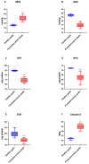
Paratuberculosis, caused by Mycobacterium avium subspecies paratuberculosis (MAP), is a chronic granulomatous enteritis with significant implications for ruminant health, economic productivity, and potential zoonotic risk. This study investigated the expression of biomarkers of oxidative stress and apoptosis in goats naturally infected with MAP, focusing on three biological matrices: serum, intestinal mucosa, and mesenteric lymph nodes. Twenty MAP-positive goats and ten healthy controls were included. Serum and tissue levels of malondialdehyde (MDA), glutathione S-transferase (GST), glutathione peroxidase (GPX), superoxide dismutase (SOD), glutathione reductase (GSR), and caspase-3 were quantitatively assessed using ELISA tests. Gross and histopathological analyses confirmed MAP infection. Infected animals showed significantly elevated serum levels of MDA and caspase-3 (p < 0.001), along with decreased antioxidant enzyme activities (GSR, GST, GPX, SOD). Tissue analysis revealed increased MDA and caspase-3 levels, particularly in the intestinal mucosa compared to mesenteric lymph nodes, suggesting localized oxidative damage and apoptosis. Conversely, antioxidant enzyme activity was higher in mesenteric lymph nodes, indicating a compensatory response and a pronounced involvement of the intestinal tract. These findings demonstrate that MAP infection induces marked oxidative stress and apoptotic processes, especially in the intestinal mucosa. The imbalance between pro-oxidant and antioxidant systems may play a key role in the pathogenesis and chronic progression of the disease. Caspase-3 and MDA, in particular, have been identified as promising diagnostic or prognostic biomarkers for MAP infection. This study highlights the importance of developing improved diagnostic tools and therapeutic strategies targeting oxidative stress pathways in paratuberculosis.
Keywords: Johne’s disease; Mycobacterium avium subspecies paratuberculosis; antioxidants; apoptosis; oxidative stress.
Conflict of interest statement
The authors declare no conflicts of interest.
Figures


References
-
- Agrawal G., Aitken J., Hamblin H., Collins M., Borody T.J. Putting Crohn’s on the MAP: Five Common Questions on the Contribution of Mycobacterium avium Subspecies Paratuberculosis to the Pathophysiology of Crohn’s Disease. Dig. Dis. Sci. 2021;66:348–358. doi: 10.1007/s10620-020-06653-0. - DOI - PMC - PubMed
-
- Navarro León A.I., Muñoz M., Iglesias N., Blanco-Vázquez C., Balseiro A., Milhano Santos F., Ciordia S., Corrales F.J., Iglesias T., Casais R. Proteomic Serum Profiling of Holstein Friesian Cows with Different Pathological Forms of Bovine Paratuberculosis Reveals Changes in the Acute-Phase Response and Lipid Metabolism. J. Proteome Res. 2024;23:2762–2779. doi: 10.1021/acs.jproteome.3c00244. - DOI - PMC - PubMed
MeSH terms
Substances
LinkOut - more resources
Full Text Sources
Research Materials

