. 2025 May 30;17(6):273.
doi: 10.3390/toxins17060273.

The Role of a Conserved Arg-Asp Pair in the Structure and Function of Tetanus Neurotoxin

Affiliations

The Role of a Conserved Arg-Asp Pair in the Structure and Function of Tetanus Neurotoxin

Elizabeth A Wilson et al. Toxins (Basel). .

Abstract

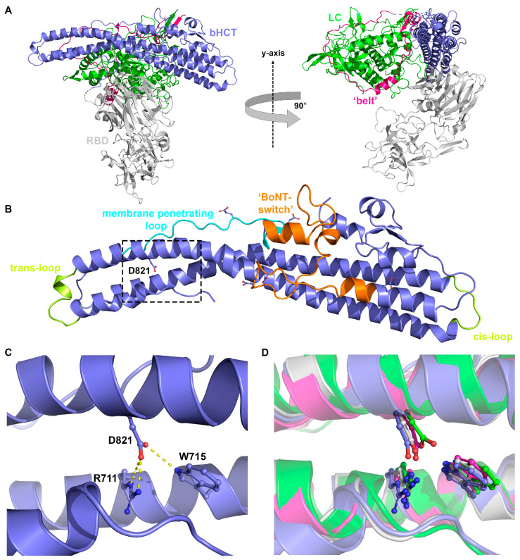
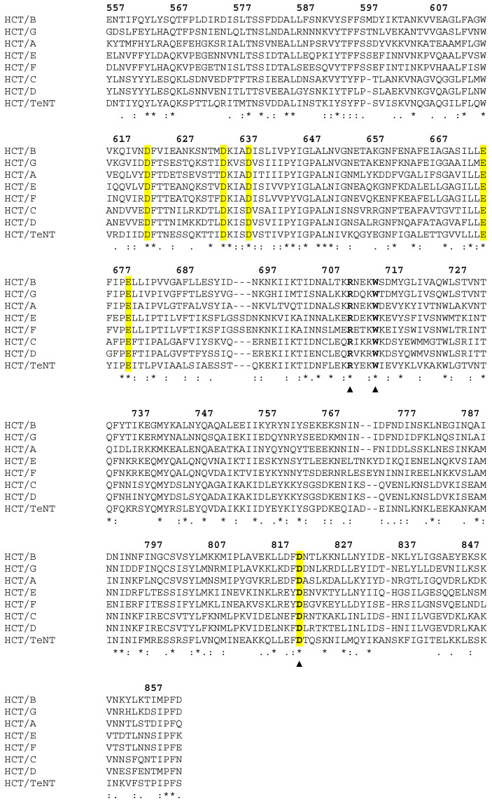
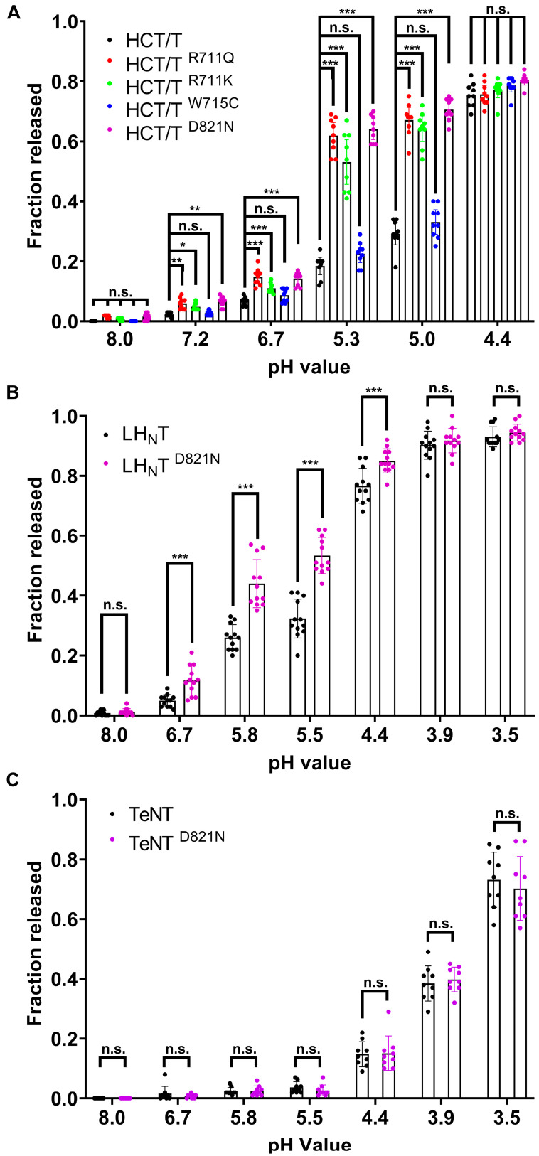
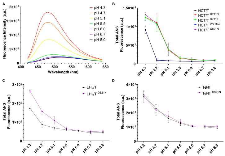
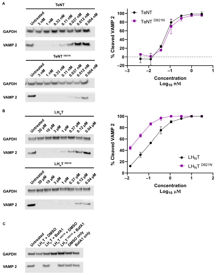
Tetanus, a severe and life-threatening illness caused by Clostridium tetani, produces symptoms such as muscle spasms, muscle stiffness and seizures caused by the production of tetanus neurotoxin (TeNT). TeNT causes spastic paralysis through the inhibition of neurotransmission in spinal inhibitory interneurons. This is achieved, in part, through pH-triggered membrane insertion of the translocation (HCT) domain, which delivers the catalytic light-chain (LC) domain to the cytosol. While the function of HCT is well defined, the mechanism by which it accomplishes this task is largely unknown. Based on the crystal structure of tetanus neurotoxin, we identified potential polar interactions between arginine 711, tryptophan 715 and aspartate 821 that appear to be evolutionarily conserved across the clostridial neurotoxin family. We show that the disruption of the Asp-Arg pair in a beltless HCT variant (bHCT) results in changes in thermal stability without significant alterations to the overall secondary structure. ANS (1-anilino-8-napthalene sulfonate) binding studies, in conjunction with liposome permeabilization assays, demonstrate that mutations at R711 or D821 trigger interactions with the membrane at higher pH values compared to wildtype bHCT. Interestingly, we show that the introduction of the D821N mutation into LHNT (LC-HCT only), but not the holotoxin, resulted in the increased cleavage of VAMP 2 in cortical neurons relative to the wildtype protein. This suggests that, as observed for botulinum toxin A, the receptor-binding domain is not necessary for LC translocation but rather helps determine the pH threshold of membrane insertion. The mutation of W715 did not result in detectable changes in the activity of either bHCT or the holotoxin, suggesting that it plays only a minor role in stabilizing the structure of the toxin. We conclude that the protonation of D821 at low pH disrupts interactions with R711 and W715, helping to drive the conformational refolding of HCT needed for membrane insertion and the subsequent translocation of the LC.

Keywords: clostridial neurotoxins; pH trigger; pore formation; tetanus toxin; translocation.


Conflict of interest statement

The authors declare no conflicts of interest.

Figures

Figure 1
Figure 1
(A) Overall structure of TeNT (PDB: 5N0B) with the catalytic domain (LC, green), the belt region (pink), the beltless translocation domain (bHCT, blue) and the receptor-binding domain (RBD, silver). (B) Enlarged view of the beltless translocation domain highlighting secondary structures implicated in LC translocation: “BoNT-switch” (orange); membrane-penetrating loop (cyan); trans- and cis-loops (yellow). In addition, the six D/E residues in TeNT conserved across the CNT family are shown as sticks (atomic color). The boxed region containing D821 is expanded in panel C. (C) Details of the interface between the two central α-helices (blue): residues Arg711-Trp715-Asp821 involved in the interface are shown as sticks (atomic color). Hydrogen bonds are indicated by yellow dashed lines. (D) Superposition of the cartoon representations of the crystal structures of BoNT/A (PDB: 3BTA, green), BoNT/B (PDB: 1EPW, silver), BoNT/E (PDB: 3FFZ, pink) and TeNT (PDB: 5N0B, blue) showing the structural conservation of the Arg-Trp-Asp triad.
Figure 2
Figure 2
Multiple sequence alignment of the beltless translocation domain of botulinum neurotoxins A–G and tetanus neurotoxin using CLUSTALW. An * (asterisk) indicates positions which have a single, fully conserved residue. Conserved acidic residues (D and E) are highlighted in yellow. The Arg711-Trp715-Asp821 triad is highlighted with ▲ symbols. Numbering refers to the sequence of tetanus neurotoxin. Here, we focus on aspartate 821 as one potential “molecular switch”, involved in triggering the conformational changes in HCT necessary for insertion into the membrane bilayer. Our rationale for choosing to focus on D821 is as follows: (i) due to its central position at the interface between the two extended amphipathic α-helices of HCT (Figure 1B,C), the protonation of D821 could very likely destabilize intramolecular contacts with arginine 711 and tryptophan 715 upon acidification; (ii) D821 and the interacting residues R711 and W715 are strictly conserved across the CNT family (Figure 1D and Figure 2); and (iii) D821 is located within a region of the central α-helix previously shown in BoNT/A to be protease-resistant in the context of proteoliposomes [21], suggesting insertion into or close association with the membrane bilayer.
Figure 3
Figure 3
Leakage of ANTS/DPX from liposomes in response to wildtype and mutated tetanus toxin proteins. The leakage of ANTS/DPX probes from 65POPC:25POPS:10Chol liposomes was measured after 15 min exposure to wildtype or mutated HCT (A), LHNT (B) and full-length toxins (C) at the indicated pH. The fluorescence (at 530 nm) of undisturbed LUVs was set to 0% and that of vesicles disrupted by Triton X-100 was set to 100%. Results are expressed as mean values ± S.D. (error bars) for a minimum of 9 independent liposome preparations. *, p < 0.05; **, p < 0.001; ***, p < 0.0001; n.s., not significant. Two-way analysis of variance with Tukey’s Multiple Comparison Test.
Figure 4
Figure 4
Tetanus toxin thermal denaturation monitored by CD spectroscopy. Average of four far-UV CD spectra of wildtype or mutated HCT (A), LHNT (C) and TeNT (E) recorded at 24 °C. In all cases, final protein concentration was 2.5 µM diluted in phosphate buffer at pH 8.0. The CD data are presented as mean residue molar ellipticity ([θ] MRW). Changes in ellipticity at 222 nm with the increase in temperature for wildtype or mutated HCT (B), LHNT (D) and TeNT (F). Data were fit to a two-state equilibrium unfolding model to estimate the thermal denaturation temperature (Tm) of each sample. Data are representative of measurements made with at least 2 independent protein preparations.
Figure 5
Figure 5
ANS binding to tetanus toxin. (A) ANS fluorescence spectra in the presence of wildtype HCT at the indicated pH values. Protein concentration was 2.5 μM, and concentration of ANS was 100 µM. Each spectrum is the average of three individual scans. Total ANS fluorescence was determined by calculating the area enclosed by the emission spectrum curve and plotted at the pH value for wildtype or mutated HCT (B), LHNT (C) and TeNT (D). Results are expressed as arithmetic mean ± SD from 3 independent determinations.
Figure 6
Figure 6
Cleavage of VAMP 2 by wildtype and mutant tetanus toxins. Cells were incubated in the presence of the indicated doses of either wildtype or mutant D821N tetanus toxins (A) or LHNTs (B). After 16 h of incubation, cells were lysed. Lysates were subjected to Western blotting with antibodies against VAMP 2 and GAPDH (loading control). Images are representative of three independent experiments. Quantification was performed using ImageJ version 1.54p. The relative cleavage of VAMP 2 was calculated by normalizing the quantification values of VAMP 2 band intensity to the total GAPDH signal. (C) Cells were incubated for 30 min with either 400 nM bafilomycin A1 (BafA1) or solvent (DMSO) prior to the addition of 30 µM LHNT or LHNT D821N. VAMP 2 cleavage was visualized and quantified as described above.
Figure 7
Figure 7
Leakage of ANTS/DPX from liposomes in response to wildtype and mutated tetanus toxin proteins. (A) The leakage of ANTS/DPX probes from 65POPC:25POPS:10Chol liposomes was measured after 15 min exposure to wildtype or mutated bHCT at the indicated pH. The fluorescence (at 530 nm) of undisturbed liposomes was set to 0% and that of vesicles disrupted by Triton X-100 was set to 100%. Results are expressed as mean values ± S.D. (error bars) for a minimum of 9 independent liposome preparations. *, p < 0.05; ***, p < 0.001; ****, p < 0.0001; n.s., not significant. Two-way analysis of variance with Tukey’s Multiple Comparison Test. (B) Average of four far-UV CD spectra of wildtype or mutated bHCT recorded at 24 °C. Final protein concentration was 2.5 µM diluted in phosphate buffer at pH 8.0. The CD data are presented as mean residue molar ellipticity ([θ] MRW).

Similar articles

References

    1. Udwadia F.E. Tetanus. Oxford University Press; Bombay, India: 1994. pp. 1–172.
    1. Rossetto O., Montecucco C. Tables of Toxicity of Botulinum and Tetanus Neurotoxins. Toxins. 2019;11:686. doi: 10.3390/toxins11120686. - DOI - PMC - PubMed
    1. Gill D.M. Bacterial toxins: A table of lethal amounts. Microbiol. Rev. 1982;46:86–94. doi: 10.1128/mr.46.1.86-94.1982. - DOI - PMC - PubMed
    1. Schiavo G., Matteoli M., Montecucco C. Neurotoxins affecting neuroexocytosis. Physiol. Rev. 2000;80:717–766. doi: 10.1152/physrev.2000.80.2.717. - DOI - PubMed
    1. Rossetto O., Pirazzini M., Montecucco C. Botulinum neurotoxins: Genetic, structural and mechanistic insights. Nat. Rev. Microbiol. 2014;12:535–549. doi: 10.1038/nrmicro3295. - DOI - PubMed

LinkOut - more resources