

Role of the IL-6/STAT3 Signaling Axis in the Protective Effect of Selenomethionine Against Zearalenone-Induced Hepatic Inflammatory Injury in Rabbits
- PMID: 40559853
- PMCID: PMC12197447
- DOI: 10.3390/toxins17060275
Role of the IL-6/STAT3 Signaling Axis in the Protective Effect of Selenomethionine Against Zearalenone-Induced Hepatic Inflammatory Injury in Rabbits
Abstract
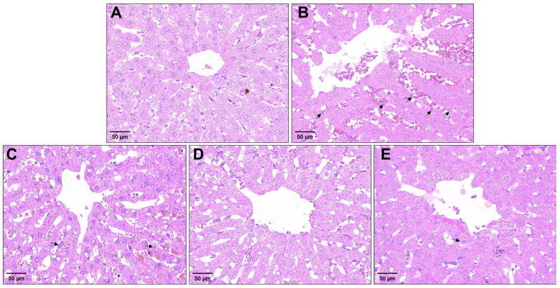
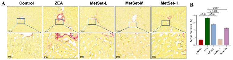
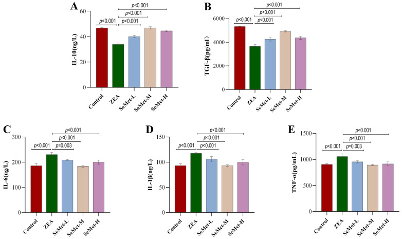
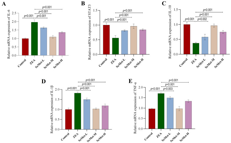
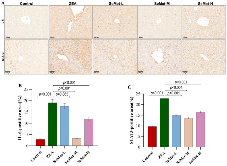
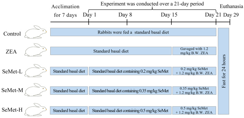
Zearalenone (ZEA), a mycotoxin primarily generated by the Fusarium species, constitutes a prevalent contaminant in both human and animal feedstuffs. Chronic exposure to this mycotoxin induces hepatic inflammatory responses in livestock species including rabbits, ultimately leading to organ damage. Selenomethionine (SeMet), an organic selenium source recognized for its antioxidant properties and anti-inflammatory bioactivity, demonstrates protective benefits in animals through its detoxification mechanism and growth promotion. The present study investigated the protective effect of SeMet against ZEA-induced hepatic inflammation and elucidated its underlying mechanisms. Fifty healthy 90-day-old rabbits were randomly divided into five groups: control, ZEA-exposed and three SeMet-supplemented groups receiving 0.2, 0.35 or 0.5 mg/kg via dietary inclusion. After two weeks of SeMet pretreatment, ZEA administration (1.2 mg/kg B.W.) was imitated via oral gavage daily for one week in both the ZEA group and three SeMet-treated groups. As a result, ZEA exposure induced the significant structural disruption of the hepatic lobules, accompanied by increased collagen deposition, elevated pro-inflammatory cytokine profiles (IL-6, IL-1β, TNF-α) and reduced anti-inflammatory mediator levels (IL-10, TGF-β). SeMet supplementation alleviated ZEA-induced histological alterations, including inflammatory cell infiltration and collagen accumulation. Biochemical analysis indicated the restoration of inflammatory markers to near-normal levels when treated with SeMet. Notably, immunohistochemical results showed that SeMet significantly reduced the protein levels of IL-6 and its downstream target STAT3 under ZEA exposure. These findings indicated that SeMet attenuated ZEA-induced hepatic inflammation by modulating the IL-6/STAT3 signaling axis, with dietary supplementation of 0.35 mg/kg SeMet exhibiting the most significant effect on alleviating ZEA-induced hepatic inflammatory injury.
Keywords: IL-6/STAT3 signaling axis; hepatic injury; inflammatory cytokines; selenomethionine; zearalenone.
Conflict of interest statement
The authors declare that there are no conflicts of interest.
Figures
References
-
- Pinton P., Tsybulskyy D., Lucioli J., Laffitte J., Callu P., Lyazhri F., Grosjean F., Bracarense A.P., Kolf-Clauw M., Oswald I.P. Toxicity of deoxynivalenol and its acetylated derivatives on the intestine: Differential effects on morphology, barrier function, tight junction proteins, and mitogen-activated protein kinases. Toxicol. Sci. 2012;130:180–190. doi: 10.1093/toxsci/kfs239. - DOI - PubMed
-
- Pistol G.C., Braicu C., Motiu M., Gras M.A., Marin D.E., Stancu M., Calin L., Israel-Roming F., Berindan-Neagoe I., Taranu I. Zearalenone mycotoxin affects immune mediators, MAPK signalling molecules, nuclear receptors and genome-wide gene expression in pig spleen. PLoS ONE. 2015;10:e0127503. doi: 10.1371/journal.pone.0127503. - DOI - PMC - PubMed
MeSH terms
Substances
Grants and funding
LinkOut - more resources
Full Text Sources
Medical
Research Materials
Miscellaneous
