

The Elias University Hospital Approach: A Visual Guide to Ultrasound-Guided Botulinum Toxin Injection in Spasticity: Part II-Proximal Upper Limb Muscles
- PMID: 40559854
- PMCID: PMC12197687
- DOI: 10.3390/toxins17060276
The Elias University Hospital Approach: A Visual Guide to Ultrasound-Guided Botulinum Toxin Injection in Spasticity: Part II-Proximal Upper Limb Muscles
Abstract
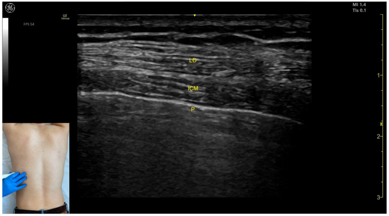
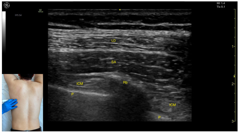
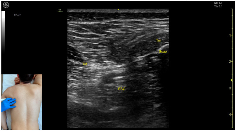
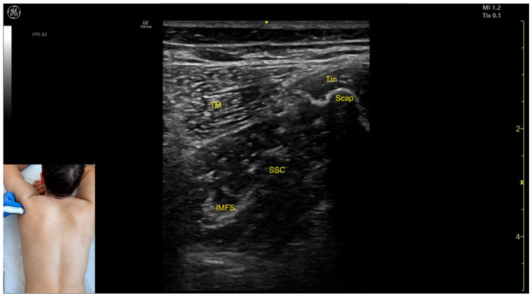
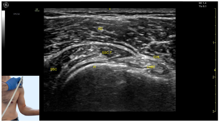
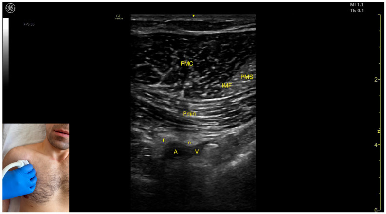
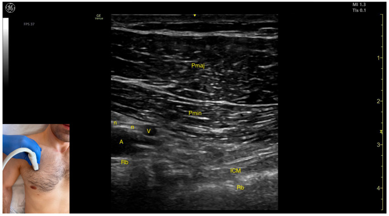
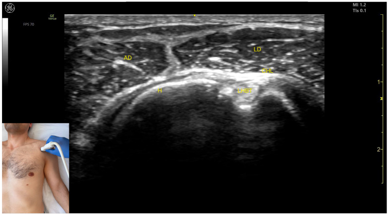
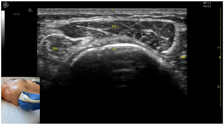
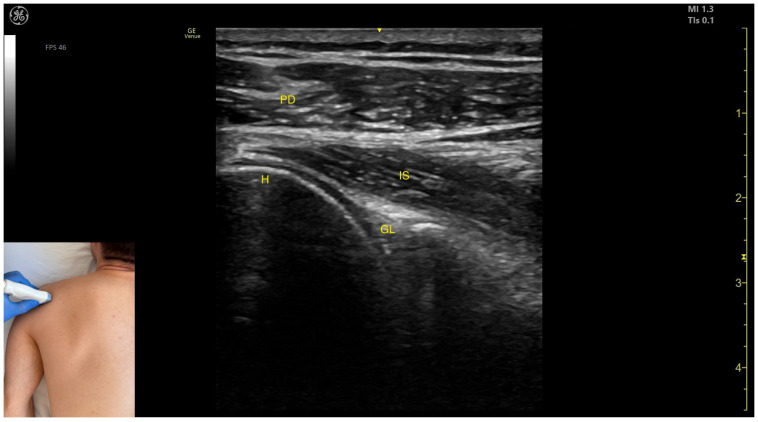
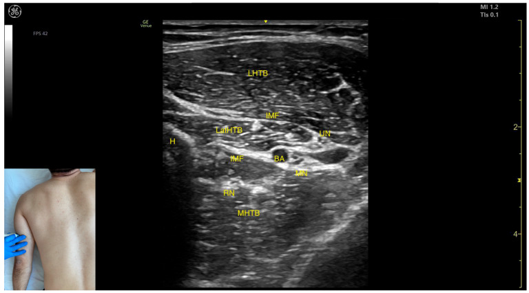
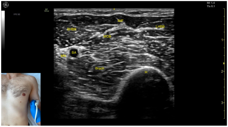
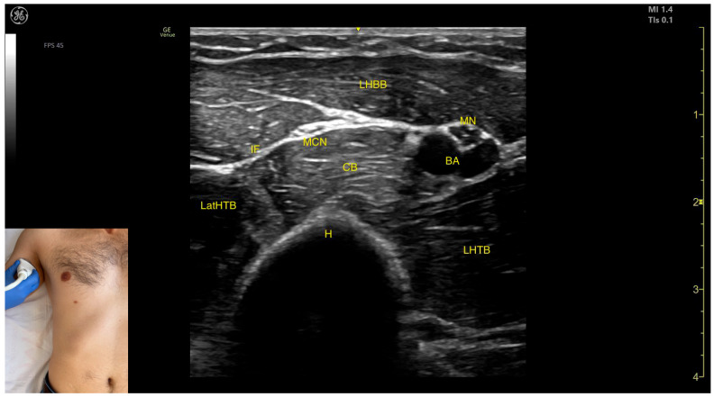
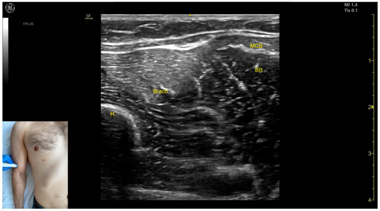
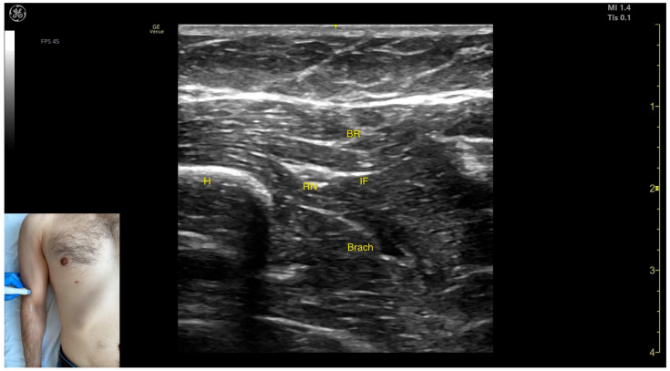
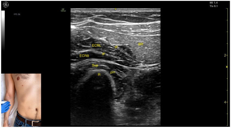
Ultrasound-guided botulinum toxin type A (BoNT-A) injections play a critical role in the management of upper limb spasticity. This is the second part of 'The Elias University Hospital Approach: A Visual Guide to Ultrasound-Guided Botulinum Toxin Injection in Spasticity' and it focuses on the proximal upper limb muscles, complementing the first part, which addressed the distal upper limb muscles. This guide provides a detailed analysis of ultrasound anatomy, clinical relevance, and injection strategies for the latissimus dorsi, teres major, subscapularis, pectoralis major, pectoralis minor, deltoid, triceps brachii, biceps brachii, brachialis, and brachioradialis. Using the Elias University Hospital (EUH) model, it presents a structured approach to BoNT-A administration, ensuring precision, safety, and optimal outcomes in spasticity management. To enhance clinical application, this guide incorporates a wide array of high-quality ultrasound images and dynamic videos, offering a comprehensive and practical understanding of scanning techniques, anatomical structures, and injection procedures. This second part of the series serves as an essential reference for clinicians, aligning with the first installment to provide a complete and systematic approach to ultrasound-guided BoNT-A therapy for upper limb spasticity.
Keywords: botulinum toxin-A injections; musculoskeletal ultrasound; post-stroke spasticity; proximal upper limb muscles; ultrasound-guided therapy.
Conflict of interest statement
The authors declare no conflicts of interest.
Figures
Similar articles
-
The Elias University Hospital Approach: A Visual Guide to Ultrasound-Guided Botulinum Toxin Injection in Spasticity: Part I-Distal Upper Limb Muscles.Toxins (Basel). 2025 Feb 26;17(3):107. doi: 10.3390/toxins17030107. Toxins (Basel). 2025. PMID: 40137880 Free PMC article. Review.
-
The Elias University Hospital Approach: A Visual Guide to Ultrasound-Guided Botulinum Toxin Injection in Spasticity: Part III-Proximal Lower Limb Muscles.Toxins (Basel). 2025 May 13;17(5):240. doi: 10.3390/toxins17050240. Toxins (Basel). 2025. PMID: 40423325 Free PMC article. Review.
-
Ultrasound Assessment of Muscle and Fascia Properties and the Effects of Spasticity and Botulinum Toxin Injections on Spastic Muscles in Chronic Stroke Survivors.Am J Phys Med Rehabil. 2025 Jun 1;104(6):527-533. doi: 10.1097/PHM.0000000000002660. Epub 2024 Nov 14. Am J Phys Med Rehabil. 2025. PMID: 39774342
-
Efficacy and safety of botulinum toxin type A for upper limb spasticity after stroke or traumatic brain injury: a systematic review with meta-analysis and trial sequential analysis.Eur J Phys Rehabil Med. 2017 Apr;53(2):256-267. doi: 10.23736/S1973-9087.16.04329-X. Epub 2016 Nov 11. Eur J Phys Rehabil Med. 2017. PMID: 27834471
-
Ultrasound visualization of Botulinum toxin injection: Identifying clinical correlates to optimize outcomes in spasticity management.PM R. 2025 Mar;17(3):254-261. doi: 10.1002/pmrj.13269. Epub 2024 Oct 22. PM R. 2025. PMID: 39436090
References
-
- Popescu M.N., Petca R.C., Beiu C., Dumitrascu M.C., Petca A., Mehedintu C., Farcasanu P.D., Sandru F. Efficiency of Different Preparations of Botulinum Toxin Type A, Xeomin and Dysport, in the Management of Spastic Upper Limb After Stroke. Rehabilitation. 2019;70:3490–3494. doi: 10.37358/RC.19.10.7582. - DOI
-
- Specchia A., Ferreri G., Cassetta E., Cerbarano L., Bentivoglio A.R. Practical Handbook for Ultrasound-Guided Botulinum Toxin Injection. Edi.Ermes srl; Millan, Italy: 2020.
-
- Kara M., Kaymak B., Ulaşli A.M., Tok F., Öztürk G.T., Chang K.-V., Hsiao M.-Y., Hung C.-Y., Yağiz On A., Özçakar L. Sonographic Guide for Botulinum Toxin Injections of the Upper Limb: EUROMUSCULUS/USPRM Spasticity Approach. Eur. J. Phys. Rehabil. Med. 2018;54:469–485. doi: 10.23736/S1973-9087.17.04664-0. - DOI - PubMed
Publication types
MeSH terms
Substances
LinkOut - more resources
Full Text Sources
Medical
