. 2025 Jun 25;74(8):251.
doi: 10.1007/s00262-025-04110-3.

Tumor-promoting function of paternally expressed gene 10 and its immunogenicity in inducing anti-tumor helper T cells in head and neck squamous cell carcinoma

Affiliations

Tumor-promoting function of paternally expressed gene 10 and its immunogenicity in inducing anti-tumor helper T cells in head and neck squamous cell carcinoma

Hiroki Komatsuda et al. Cancer Immunol Immunother. .

Abstract

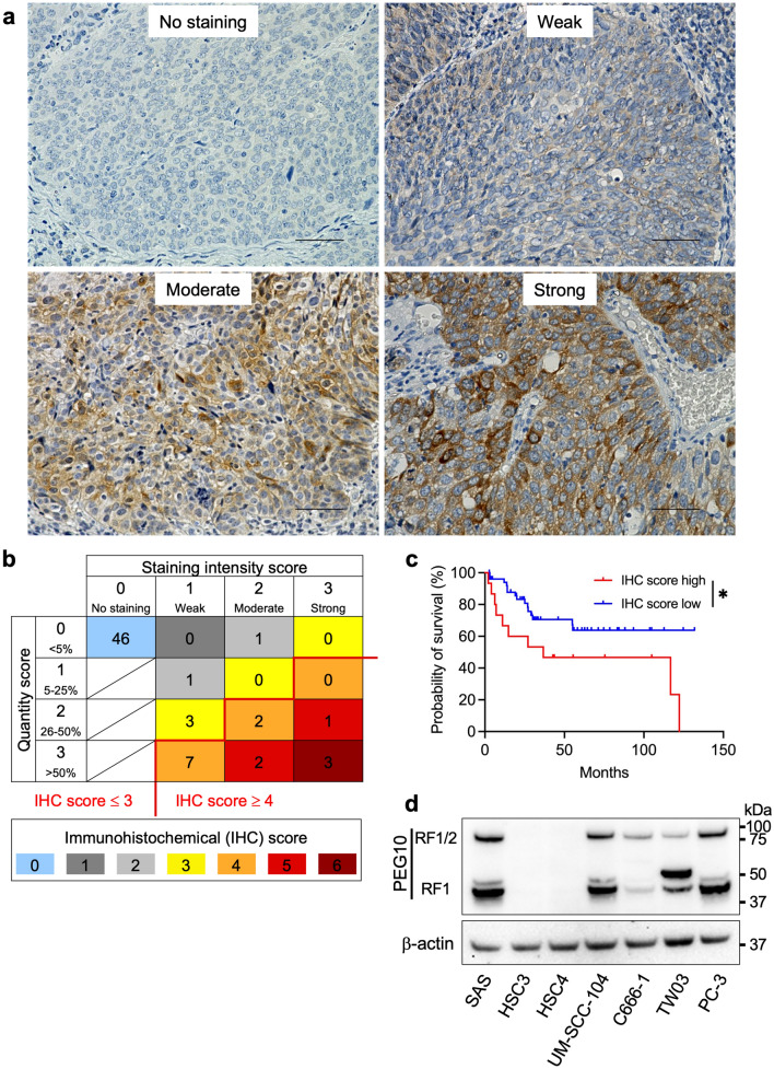
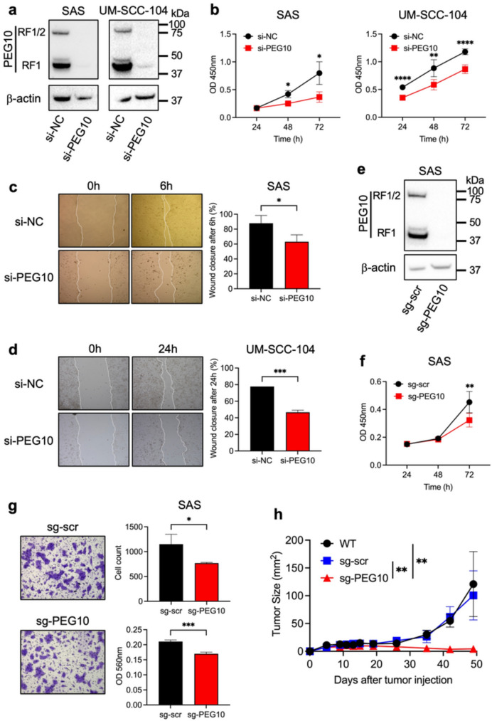
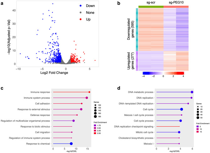
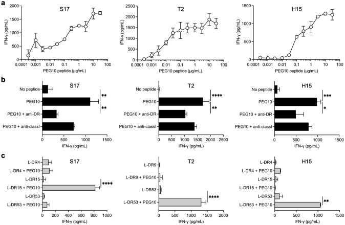
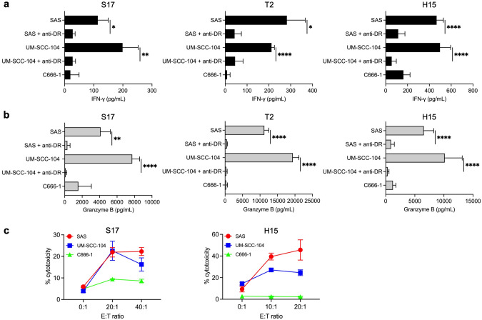
Paternally expressed gene 10 (PEG10) is expressed primarily in the placenta; its expression is extremely low or absent in normal tissues but up-regulated in various cancers, indicating that PEG10 is a potential target for cancer immunotherapy. However, the expression and role of PEG10 in head and neck squamous cell carcinoma (HNSCC) and the immunogenicity of possible PEG10-derived T-cell epitopes remain unclear. In the present study, we show that PEG10 is expressed in HNSCC, and its high expression is associated with poor patient survival. Suppression of PEG10 expression attenuated the proliferation, migration, and invasion of HNSCC cells and altered their gene expression profiles. We also identified a PEG10-derived peptide epitope (PEG10216-232) capable of eliciting antigen-specific and promiscuous human leukocyte antigen (HLA)-DR-restricted helper T lymphocyte (HTL) responses. Notably, PEG10-specific HTLs exerted direct cytotoxicity against PEG10-positive HNSCC cells in an HLA-DR-restricted manner. Moreover, precursor T cells that react to PEG10216-232 peptide were detected in HNSCC patients. These results indicate that PEG10 plays an important role in HNSCC tumorigenesis and qualifies as an immunotherapeutic target against HNSCC. The helper epitope peptide of PEG10 could effectively stimulate antigen-specific HTLs and induce anti-tumor responses against PEG10-positive cancers, including HNSCC.

Keywords: Epitope; Head and neck squamous cell carcinoma; Helper T lymphocytes; Paternally expressed gene 10; Peptide vaccine; Placenta.


Conflict of interest statement

Declarations. Conflict of interest: The authors declare no competing interests. Ethical approval: This study was performed in compliance with the principles of the Declaration of Helsinki and subsequent amendments, and the study protocol was approved by the Institutional Review Board of the Asahikawa Medical University (#15005 and #16217). All animal experiments were approved by the Institutional Animal Care and Use Committee of Asahikawa Medical University (R6-035). Informed consent: As this was a retrospective study using existing specimens and clinical information, the Institutional Review Board approved a waiver of the standard informed consent process for IHC analyses. Instead, an opt-out approach was implemented through the Asahikawa Medical University website that ensured patients retained the right to decline participation in the study. All patients from whom PBMCs were collected received a written explanation of the study and provided written informed consent before participation.

Figures

Fig. 1
Fig. 1
Expression of PEG10 in HNSCC patients and cell lines. a Representative IHC staining images of PEG10 in oropharyngeal SCC specimens. Expression levels in tumor cells were scored as no, weak, moderate, or strong IHC staining intensity. Scale bars = 50 μm. b Distribution of IHC scores for PEG10. The IHC score was calculated as the sum of the staining intensity score (0, no staining; 1, weak; 2, moderate; 3, strong) and quantity score (percentage of positively stained tumor cells: 0, < 5%; 1, 5–25%; 2, 26–50%; 3, > 50%). The IHC score could range from 0 to 6. An IHC score ≥ 4 was defined as high expression, and lower scores were defined as indicating low expression. c Kaplan–Meier curves for disease-specific survival of oropharyngeal SCC patients divided into two groups based on PEG10 IHC score. Asterisk indicates statistical significance at P < 0.05. d Western blot analysis of PEG10 expression in tumor cell lines. β-actin was used as a loading control. PC-3 cells were used as a positive control for PEG10 expression
Fig. 2
Fig. 2
Effect of PEG10 suppression in HNSCC cells. a Western blot analysis of PEG10 expression in SAS and UM-SCC-104 cells transfected with negative control siRNA (si-NC) or PEG10 siRNA (si-PEG10). β-actin was used as a loading control. b CCK-8 assay of the proliferation of SAS and UM-SCC-104 cells transfected with si-NC or si-PEG10 at 24 h, 48 h, and 72 h. c, d Wound healing assay of the migration of SAS (c) and UM-SCC-104 (d) cells transfected with si-NC or si-PEG10. Representative images at indicated timepoints (left panels) and quantification (right panels) of wound closure are shown. e Western blot analysis of PEG10 expression in SAS cells transfected with non-targeting scrambled sgRNA (sg-scr) or PEG10 sgRNA (sg-PEG10). β-actin was used as a loading control. f CCK-8 assay of the proliferation of SAS cells transfected with sg-scr or sg-PEG10 at 24 h, 48 h, and 72 h. g Assay using transwells coated with extracellular matrix gel to assess the invasion of SAS cells transfected with sg-scr or sg-PEG10. Representative images (left panels) and quantification (right panels) of invasive cells at 24 h are shown. h Tumor growth in nude mice xenografted with un-transfected wild-type (WT), sg-scr–transfected, or sg-PEG10–transfected SAS cells. In b, c, d, f, and g, data are shown as mean ± SD or + SD from at least triplicate measurements. In h, data are shown as mean ± SD of 3 mice per group. One, two, three, and four asterisks indicate statistical significance at P < 0.05, P < 0.01, P < 0.001, and P < 0.0001, respectively
Fig. 3
Fig. 3
RNA-seq analysis of SAS cells transfected with PEG10 sgRNA (sg-PEG10) or non-targeting scrambled sgRNA (sg-scr). a, b Volcano plot (a) and heatmap (b) showing differentially regulated genes in sg-PEG10 compared with sg-scr. RNA-seq analysis revealed a total of 595 downregulated genes and 277 upregulated genes. c, d Downregulated (c) and upregulated (d) GO enrichment biological processes in sg-PEG10 compared with sg-scr. Graphs display the top 10 enriched GO terms
Fig. 4
Fig. 4
Characteristics of induced PEG10-specific CD4 HTLs. a Evaluation of the peptide dose responses of PEG10216-232-specific HTLs (S17, T2, and H15). These T cells were co-cultured with autologous PBMCs pulsed with various concentrations of PEG10216-232 peptide for 48 h. Supernatants were harvested, and the secretion of IFN-γ was measured using ELISA. Data are shown as mean ± SD from at least duplicate measurements. b Determination of the HLA restriction of PEG10216-232-specific HTLs. The response of T cells co-cultured with autologous PBMCs pulsed with PEG10216-232 peptide was evaluated in the presence of anti–HLA-DR Ab or anti–HLA class I Ab (negative control). c Detailed determination of which HLA-DR molecules are responsible for PEG10216-232 peptide presentation. The response of HTLs to PEG10216-232 peptide was measured using peptide-pulsed L-cells transfected with individual HLA-DR alleles as APCs. In b and c, supernatants were harvested after 48 h, and IFN-γ secretion was measured using ELISA. Data are shown as mean + SD from at least triplicate measurements. One, two, three, and four asterisks indicate statistical significance at P < 0.05, P < 0.01, P < 0.001, and P < 0.0001, respectively
Fig. 5
Fig. 5
Direct recognition and cytotoxic activity of PEG10-specific HTLs against HLA-DR–matched tumor cells expressing PEG10. a, b To evaluate the capacity to recognize naturally processed PEG10 antigen in tumor cells, PEG10216-232-specific HTLs (DR15-restricted T cells, S17; DR53-restricted T cells, T2 and H15) were co-cultured with HLA-DR15 and DR53-positive (SAS and UM-SCC-104) and HLA-DR–unmatched (C666-1) tumor cells expressing PEG10 with/without anti–HLA-DR Ab. Supernatants were harvested and subjected to ELISA for IFN-γ (a) and granzyme B (b) release after 48 h of co-culture. c S17 or H15 cells were co-cultured with CFSE-labeled SAS, UM-SCC-104, and C666-1 cells at various effector:target (E:T) ratios. After 6 h of co-culture, the cells were collected to determine the percentage of CFSE-labeled dead cells using flow cytometry with 7-AAD. In ac, C666-1 cells were used as negative controls. Data are shown as mean + SD or ± SD from at least triplicate measurements. One, two, and four asterisks indicate statistical significance at P < 0.05, P < 0.01, and P < 0.0001, respectively
Fig. 6
Fig. 6
Evaluation of T-cell responses to PEG10 peptide in HNSCC patients. The characteristics of the 6 HNSCC patients are summarized in the upper table. PBMCs derived from HNSCC patients were cultured in the presence of PEG10216-232 or PADRE peptide for the first 7 days and restimulated with peptide-pulsed irradiated autologous PBMCs for the next 7 days. PADRE peptide was used as a positive control for activating CD4 T cells. Anti–HLA-DR Ab was used to block peptide presentation by HLA-DR. Supernatants were harvested and analyzed for IFN-γ release using ELISA. Data are shown as mean + SD from at least triplicate measurements. One, two, three, and four asterisks indicate statistical significance at P < 0.05, P < 0.01, P < 0.001, and P < 0.0001, respectively

Similar articles

References

    1. Ferris RL, Blumenschein G Jr, Fayette J, Guigay J, Colevas AD, Licitra L, Harrington K, Kasper S, Vokes EE, Even C, Worden F, Saba NF, Iglesias Docampo LC, Haddad R, Rordorf T, Kiyota N, Tahara M, Monga M, Lynch M, Geese WJ, Kopit J, Shaw JW, Gillison ML (2016) Nivolumab for recurrent squamous-cell carcinoma of the head and neck. N Engl J Med 375:1856–1867. 10.1056/NEJMoa1602252 - PMC - PubMed
    1. Hirayama M, Nishimura Y (2016) The present status and future prospects of peptide-based cancer vaccines. Int Immunol 28:319–328. 10.1093/intimm/dxw027 - PubMed
    1. Kumai T, Kobayashi H, Harabuchi Y, Celis E (2017) Peptide vaccines in cancer-old concept revisited. Curr Opin Immunol 45:1–7. 10.1016/j.coi.2016.11.001 - PMC - PubMed
    1. Quezada SA, Simpson TR, Peggs KS, Merghoub T, Vider J, Fan X, Blasberg R, Yagita H, Muranski P, Antony PA, Restifo NP, Allison JP (2010) Tumor-reactive CD4(+) T cells develop cytotoxic activity and eradicate large established melanoma after transfer into lymphopenic hosts. J Exp Med 207:637–650. 10.1084/jem.20091918 - PMC - PubMed
    1. Oh DY, Kwek SS, Raju SS, Li T, McCarthy E, Chow E, Aran D, Ilano A, Pai CS, Rancan C, Allaire K, Burra A, Sun Y, Spitzer MH, Mangul S, Porten S, Meng MV, Friedlander TW, Ye CJ, Fong L (2020) Intratumoral CD4(+) T cells mediate anti-tumor cytotoxicity in human bladder cancer. Cell 181(1612–1625):e1613. 10.1016/j.cell.2020.05.017 - PMC - PubMed

MeSH terms