. 2025 Jun 25;23(6):e3003223.
doi: 10.1371/journal.pbio.3003223. eCollection 2025 Jun.

Ancestral [Fe-S] biogenesis system SMS has a unique mechanism of cluster assembly and sulfur utilization

Affiliations

Ancestral [Fe-S] biogenesis system SMS has a unique mechanism of cluster assembly and sulfur utilization

Macha Dussouchaud et al. PLoS Biol. .

Abstract

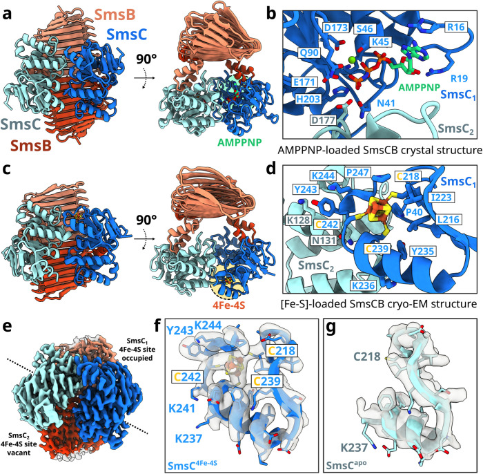
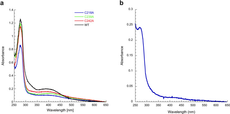
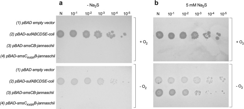
[Fe-S] clusters are ancient and ubiquitous protein co-factors, which contributed to the emergence of life in an anoxic planet. We have recently identified two minimal [Fe-S] biogenesis systems, MIS and SMS, inferred to be ancestral systems dating back to the Last Universal Common Ancestor and which gave rise to the well-studied modern Iron-Sulfur Cluster (ISC), Nitrogen Fixation (NIF), and Sulfur Mobilization (SUF) machineries. The present study focuses on the ancestor SMS from the hyperthermophilic archaeon Methanocaldococcus jannaschii. Biochemical and structural studies showed that SMS is made of a SmsC2B2 heterotetratmer wherein the SmsC subunit hosts both ATP and [Fe-S] cluster binding sites. Binding of ATP and assembly of [Fe-S] were found to be mutually exclusive allowing for a regulatory coupling between binding of both substrates. Mutagenesis and in vitro transfer experiments revealed the key role of SmsC-contained Cys residues in cluster assembly. Strikingly, the SMS system rescued a non-viable Escherichia coli strain lacking endogenous ISC and SUF systems grown under anoxic conditions, in the presence of Na2S, indicating that sulfide is a source of sulfur for SMS. In addition, we predict that most archaea SmsC proteins hold a similar C-terminal [Fe-S] cluster assembly site. Taking into account those unique structural and functional features, we propose a mechanistic model describing how SmsC2B2 assembles and distributes [4Fe-4S] clusters. Altogether this study established SMS as a new bona fide [Fe-S] biogenesis system that operated in anaerobic prokaryotes prior to evolve to SUF after the Great Oxydation Event.


Conflict of interest statement

The authors have declared that no competing interests exist.

Figures

Fig 1
Fig 1. Spectroscopy analysis of SmsC2B2 complex.
(a) SDS-PAGE and size exclusion chromatography profile of purified Sms proteins. Lane (1): V0, lane (2): SmsC2B2, lane (3): SmsC from the SmsC2B2, lane (4): SmsC monomers, lane (5): SmsB homodimers. (b) UV-Vis absorption spectrum of SmsC2B2. SmsC2B2 (38.5 μM) was incubated with 5 equivalents of Fe2+/SmsC2B2, 5 equivalents of Na2S/SmsC2B2, and 3 mM DTT. (c) 6 K Mössbauer spectra (black vertical bars) of SmsC2B2 (350 µM, 3.6 Fe and 3.4 S/SmsC2B2) recorded using a 0.06 T and a 7 T external magnetic field applied parallel to the γ-beam. The simulations assuming a unique iron site are overlaid as thick red solid lines (see text for parameters). (d) UV-Vis absorption spectrum of chemically reconstituted Sms proteins. SmsC (in red) (70 μM) and SmsB (in blue) (62 μM) were incubated with 5 equivalents of Fe2+/SmsC2 or SmsB2, 5 equivalents of Na2S/SmsC2 or SmsB2 and 3 mM DTT. The data underlying this figure can be found in Fig 1 and S1 Data.
Fig 2
Fig 2. Overall architecture of the SmsC2B2 complex.
(a) AMP-PNP-loaded SmsC2B2 crystal structure. (b) Close-up on the binding site of the AMP-PNP/Mg2+ showing residues surrounding the binding site of AMP-PNP. (c) Cryo-EM structure of the [Fe-S]-bound SmsC2B2 complex. (d) Close-up on the binding site of the [Fe-S] cluster showing the C218, C239, and C242 coordinating the [Fe-S] cluster. The cluster binding site lies in a solvent-exposed hydrophobic pocket consisting of residues P40, L216, I223, Y235, F234, and P247. (e) The [Fe-S] cluster-bound SmsC2B2 complex exhibits asymmetry. (f) The [Fe-S] bound SmsC displays a folded C-terminal α-helix composed of residues 227–241 and a short terminal loop. (g) Close-up from the apo-SmsC-COOH region till residue 237.
Fig 3
Fig 3. Proposed mechanism of the [Fe-S] biogenesis process on the scaffold SmsC2B2.
SmsC2B2 assembles first a [Fe-S] cluster (ATP not required), on one of the SmsC subunits (a, intermediate 1). Upon interaction, the [Fe-S] cluster is transferred from SmsC2B2 to the targeted apo-protein client. Upon dissociation of the cluster, the C-terminal extension of SmsC unfolds, destabilizing the asymmetric SmsC-SmsC dimer observed in the cryo-EM structure, and allowing the SmsC2B2 complex to open to adopt a conformation like the one observed in the crystal structure (a, intermediate 2). ATP binding triggers conformational changes in the P-loop (b). The SmsC subunits rearrange again to transiently form a tight dimer as seen in the SmsC2 structure favoring ATP hydrolysis (a, intermediate 3), after which ADP and Pi leave SmsC and SmsC is reset to engage into assembling a new [Fe-S] cluster (a, intermediate 1).
Fig 4
Fig 4. Residues C219, C239, and C242 of SmsC act as ligands of the [Fe-S] cluster.
(a) SmsC (84 μM) (black), SmsCC239A (84 μM) (green), SmsCC242A (84 μM) (red), and SmsCC218A (56 μM) (blue) were incubated with 5 equivalents of Fe2+/SmsC, 5 equivalents of Na2S/SmsC, and 3 mM DTT. UV-Vis absorption spectrum of SmsC shows an absorption at 420 nm compared to the variants SmsCC218A, SmsCC239A, and SmsCC242A. (b) SmsC(C239A-C242A)2B2 (50 μM) was incubated with 5 equivalents of Fe2+/SmsC(C239A-C242A)2B2, 5 equivalents of Na2S/SmsC(C239A-C242A)2B2, and 2 mM DTT. UV-Vis absorption spectrum of the SmsC(C239A-C242A)2B2variant shows no absorbance at 420 nm (1 nmol of SmsC(C239A-C242A)2B2 contains 0.23 nmol of iron and 0.3 nmol of sulfur). The data underlying this figure can be found in Fig 4 and S2 Data.
Fig 5
Fig 5. Role of ATP binding/hydrolysis in SmsCB.
(a) SmsC (35 µM), SmsCK45R (35 µM), and SmsC2B2 (35 µM) proteins were added to 1 ml of 25 mM Hepes buffer (pH 7.6) containing 100 mM KCl, 5 mM MgSO4, 5 mM phospho-enol pyruvate, 1 mM NADH, 5 UI of PK, and 10 UI of LDH. Then, 1 mM of ATP was added to initiate the reaction at 25 °C. Specific activities were calculated using the molar extinction coefficient of 6.22 mM−1 cm−1 for NADH and the protein concentrations determined from the extinction coefficient. SmsC2B2 displays an ATPase activity 10-fold higher than SmsC. SmsCK45R displays a residual ATPase activity. (b) Equilibirum binding curve. SmsC proteins from 0 to 40 µM and SmsCK45R proteins from 0 to 25 µM were incubated with 400 nM concentration of mantATPyS in HEPES buffer 25 mM, KCl 100 mM, MgSO4 5 mM, pH 7.6. Measurements were taken using a fluorimeter (λexc 355 nm and λem 448 nm for mantATPyS). Dissociation constant was calculated using the equation y = m1 * x/(m2 + x). (c) SmsC proteins first incubated with 10 mM AMP-PCP (blue) or not (red) were purified by SEC. Elution peaks of SmsC, SmsC2, AMP-PCP Sms are indicated on top of the graph. (d) SmsC (blue) and SmsCK45R (red) proteins were incubated with 10 mM of AMP-PCP and purified by SEC. Elution peaks of SmsC, SmsC2, AMP-PCP Sms are indicated on top of the graph. The data underlying this figure can be found in Fig 5 and S3 Data.
Fig 6
Fig 6. ATP and [Fe-S] cluster binding are mutually exclusive.
(a) SmsC2B2 (84 μM) (red) and AMP-PCP-SmsC2B2 (84 μM) (blue) were incubated with 5 equivalents of Fe2+/ SmsC2B2, 5 equivalents of Na2S/ SmsC2B2, and 3 mM DTT. UV-Vis absorption spectrum of SmsC2B2 shows an absorption at 420 nm compared to the AMP-PCP-SmsC2B2 bound form. (b) Relative fluorescence measurements. SmsC2B2 (50 μM) (red) and [Fe-S]-SmsC2B2 (50 μM) (blue) were incubated with 21 μM of mantATPyS. Measurements were taken using a fluorimeter (λexc 355 nm and λem 448 nm). (c) Apo-AcnB (0.5 nmol) was incubated with reconstituted SmsC2B2 (1.74 eq.; 2.9 Fe/3.0 S per SmsC2B2) ± 1 mM ATP and 2 mM MgCl₂. Aconitase activity was measured after 1, 2, and 10 min to assess the effect of ATP on [Fe-S] cluster transfer. As a positive control (RecACnB), apo-AcnB was assayed after 30 min with 5 molar excess of Fe2+ and S2− in the presence of 500 µM DTT. The initial velocity (μM isocitrate/min) was measured in duplicate, short bars correspond to mean deviation. The data underlying this figure can be found in Fig 6 and S4 Data.
Fig 7
Fig 7. Sodium sulfide is the source of sulfur for the SmsCB complex.
Spot test assay for growth indifferent culture dilutions of E. coli. ΔiscUAΔsuf MEV carrying the empty pBAD vector (lane 1), the pBAD vector carrying the Escherichia coli sufABCSDE operon (lane 2), the pBAD vector carrying the Methanocaldococcus jannaschii smsCB operon (lane 3), and the pBAD vector carrying the M. jannaschii smsCK45RB operon (lane 4). Medium was LB supplemented with 0.2% arabinose (a) or 0.2% arabinose and 5 mM Na2S (b), in oxic or anoxic conditions.
Fig 8
Fig 8. Diversity of the C-terminal region (CTR) of SmsC.
(a) Multiple sequence alignment of SmsC sequences showing that the beta-beta-alpha region is well conserved from D212 to G240 residues. Number of cysteine residues per site in the multiple alignement of 1,387 SmsC. (b) Subsample of SmsC CTR showing the diversity in terms of cysteine arrangements.
Fig 9
Fig 9. Alphafold predictions of the structures of the Sms CTRs harboring different arrangement of Cys residues.
Predictions harbor several Cys residues close to each other in the structural models, being compatible with a [Fe-S] binding cluster capacity. Alphafold prediction of the SmsCB proteins of Clostridium tetani (Ctetani), Clostridium novyi (Cnovyi), Methanopyrus kandleri (Mkandleri), Archaeoglobus fulgidus (Afulgidus), Methanococcus voltae (Mvoltae), Dehalobacter sp (Dsp), and Syntrophus aciditrophicus (Saciditrophic). Cysteine residues are shown in red.

References

    1. Fontecave M. Iron–sulfur clusters: ever-expanding roles. Nat Chem Biol. 2006;2:171–4. - PubMed
    1. Wächtershäuser G. Groundworks for an evolutionary biochemistry: the iron-sulphur world. Prog Biophys Mol Biol. 1992;58(2):85–201. doi: 10.1016/0079-6107(92)90022-x - DOI - PubMed
    1. Imlay JA. Iron-sulphur clusters and the problem with oxygen. Mol Microbiol. 2006;59(4):1073–82. doi: 10.1111/j.1365-2958.2006.05028.x - DOI - PubMed
    1. Boyd ES, Thomas KM, Dai Y, Boyd JM, Outten FW. Interplay between oxygen and Fe-S cluster biogenesis: insights from the Suf pathway. Biochemistry. 2014;53(37):5834–47. doi: 10.1021/bi500488r - DOI - PMC - PubMed
    1. Spietz RL, Payne D, Szilagyi R, Boyd ES. Reductive biomining of pyrite by methanogens. Trends Microbiol. 2022;30(11):1072–83. doi: 10.1016/j.tim.2022.05.005 - DOI - PubMed

MeSH terms

LinkOut - more resources