. 2025 Jul;12(4):e200423.
doi: 10.1212/NXI.0000000000200423. Epub 2025 Jun 25.

Neuropathic Pain and Distinct CASPR2 Autoantibody IgG Subclasses Drive Neuronal Hyperexcitability

Affiliations

Neuropathic Pain and Distinct CASPR2 Autoantibody IgG Subclasses Drive Neuronal Hyperexcitability

Margarita Habib et al. Neurol Neuroimmunol Neuroinflamm. 2025 Jul.

Abstract

Background and objectives: Patients with autoantibodies (aAbs) against the contactin-associated protein-like 2 (CASPR2) suffer from a variety of clinical syndromes including neuropathic pain. CASPR2 is an adhesion protein of the neurexin family and part of the voltage-gated potassium channel complex (VGKC complex) in dorsal root ganglia (DRG) neurons. The pathologic mechanisms following the binding of CASPR2 aAbs and their association with pain are only partially understood. CASPR2 aAbs are mainly of the IgG4 subclass; however, previous studies have neglected subclass-dependent effects.

Methods: We investigated 49 subclassified patient serum samples positive for CASPR2 aAbs combining superresolution lattice structural illumination microscopy (SIM2) and functional readouts by calcium imaging and electrophysiologic recordings on cultured DRG neurons. CASPR2-positive patient sera subclassified in IgG4 together with at least 1 other IgG subclass (IgGX) and patients with only IgG4 were further subdivided into the pain and no pain groups.

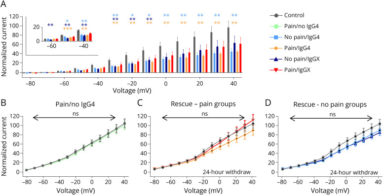
Results: A decrease of CASPR2 expression along the axons after exposure to CASPR2 aAbs was observed for all patient groups except the group without pain and IgG4. Moreover, binding of CASPR2 aAbs from patients with pain increased the distance between CASPR2 and associated potassium channels along DRG axons determined by SIM2 microscopy. CASPR2 aAbs of patients with pain significantly increased overall neuronal excitability of cultured DRG neurons as measured by calcium imaging. Patch-clamp recordings revealed significantly decreased current amplitudes of voltage-gated potassium (Kv) channels after incubation with all 4 CASPR2 aAb subclassifications with the most prominent effect of serum samples harboring IgG4 aAbs only. Replacement of patient aAbs by healthy control serum rescued Kv channel function to normal levels suggesting that the affected potassium channel function is due to structural blockage and disrupted interactions within the VGKC complex. The last might also be rescued on novel protein synthesis and membrane trafficking of CASPR2.

Discussion: IgG4 aAbs seem to be the major modifier of potassium channel function. The DRG hyperexcitability is primarily due to impaired Kv channel conductance as a consequence of CASPR2 aAb binding. However, additional unidentified signal pathways contribute to this process in patients with neuropathic pain.


Conflict of interest statement

The authors report no relevant disclosures. Full disclosure form information provided by the authors is available with the full text of this article at Neurology.org/NN.

Figures

Figure 1
Figure 1. CASPR2 aAbs in Patient Serum Show Different IgG Subclass Compositions
(A) Exemplary immunocytochemical stainings of CASPR2 (cyan) and patient serum (red) binding to membrane of CASPR2-transfected HEK-293 cells and adult DRG neurons. (B) Stainings for different IgG subclasses (cyan) in 2 exemplary total IgG positive (red) patient sera (P31, P39). (C) Distribution of IgG subclasses (IgG4 only or IgG4 plus additional IgG = IgGX) and pain phenotype with pain—red and orange circles, no pain—light and dark blue circles with circle size related to the number of positive patient sera. (D) Distribution of IgG subclass combinations in patients with pain (red) and without pain (blue). CASPR2 = Contactin-associated protein 2; DRG = dorsal root ganglia; IgG = immunoglobulin G.
Figure 2
Figure 2. Expression of CASPR2 After Long-Term Exposure to CASPR2 aAbs and Structural Integrity of the VGKC Complex
(A) Microfluidic chamber (MFC) with embryonic DRGs to separate somatic and axonal compartments and investigate VGKC complex organization after CASPR2 aAb incubation in separated compartments. (B) Exemplary pictures using lattice SIM2 microscopy of soma and axons from DRGs in MFCs treated with healthy control serum. Arrows mark the distance between CASPR2 (magenta) and Kv1.2 (green). (C) Quantification of distance between CASPR2 and Kv1.2 for somata (left) and axons (right) after CASPR2 aAb incubation for 2 hours. Data are shown as mean ± SEM. Somatic and axonal complexes: n = 325–1,465 and n = 1872–3,603, respectively. (D) CASPR2 and Kv1.2 density per 100 µm axon (CASPR2 n = 34–37; Kv1.2 n = 33–37). Data are shown as violin plots with individual values, median = bold line, quartiles = dotted lines. Levels of significance: ns: not significant, *p < 0.05, **p < 0.01, ****p < 0.0001. CASPR2 = Contactin-associated protein 2; DRG = dorsal root ganglia; VGKC complex = voltage-gated potassium channel complex.
Figure 3
Figure 3. Spontaneous Calcium Activity of DRGs After Incubation With CASPR2 aAbs
(A) Exemplary pictures and activity graphs of ROIs after short-term incubation with control serum or serum group with pain and IgGX. Neuronal activity from ∼40 cells is shown in a heatmap. (B) Frequency (left), amplitude (middle), and area under the curve (AUC; right) of spontaneous calcium activity events after short-term incubation with CASPR2 aAbs of different serum subclassifications. Data shown as mean ± SEM, n = 718 (control), n = 682 (no pain/IgG4), n = 476 (no pain/IgGX), n = 566 (pain/IgG4), and n = 727 (pain/IgGX). Levels of significance: *p < 0.05, **p < 0.01, ****p < 0.0001. aAbs = autoantibodies; CASPR2 = Contactin-associated protein 2; DRG = dorsal root ganglia.
Figure 4
Figure 4. Decrease of Kv Currents on CASPR2 aAb Application
(A) Bar plot of potassium channel activity at different voltage steps from −80 to +40 mV. Normalized current compared with the healthy control is shown. (B) I–V (current-voltage relation) plot measured after short-term aAb application on DRG neurons under the condition: pain/no IgG4, N = 2, n = 10. (C and D) Replacement of the CASPR2 aAbs by healthy control serum led to a rescue of affected potassium channel current (patients with pain and patients without pain), N = 2, n = 10–11. Levels of significance: *p < 0.05, **p < 0.01, ***p < 0.001. aAbs = autoantibodies; CASPR2 = Contactin-associated protein 2; DRG = dorsal root ganglia; Kv = voltage-gated potassium.
Figure 5
Figure 5. CASPR2 Monoclonal Autoantibodies Isolated From Patients Also Decrease Potassium Channel Activity in DRG Neurons
(A and B) Representative potassium channel current recording and the effect of specific potassium channel subunit composition blocker α-dendrotoxin Dtx-A and κM-RIIIJ (red traces) shown in the bar plot (at +40 mV). Data shown as mean ± SEM. (C) Domain architecture of CASPR2 including the discoidin domain, fibrinogen-like domain, and laminin domains. Patch clamp recordings using a voltage step protocol from −80 to +40 mV in the presence of CASPR2 monoclonal autoantibodies against the discoidin domain (left) demonstrating significant alterations of the potassium channel activity and against the laminin domain (right) with no significant alterations. N = 3, n = 13–16. Level of significance: *p < 0.05, ****p < 0.0001. CASPR2 = Contactin-associated protein 2; DRG = dorsal root ganglia.
Figure 6
Figure 6. Pathophysiologic Mechanisms of CASPR2 aAbs Following Patient Subclassification According to IgG Subtype and Pain Phenotype
DRG neurons are active mediators in developing neuropathic pain: healthy condition (left) and disease condition in the presence of CASPR2 autoantibodies (aAbs, right). Sensory neuron hyperexcitability is driven by pain-associated CASPR2 aAbs. This causes enhanced neuronal activity and decreased function of the associated Kv channels. Pathologic activity of sensory neurons is mainly promoted by CASPR2 aAbs of the IgG4 subtype. Created in BioRender. Villmann, C. (2025) BioRender.com/. CASPR2 = Contactin-associated protein 2; DRG = dorsal root ganglia.

References

    1. Dalmau J, Geis C, Graus F. Autoantibodies to synaptic receptors and neuronal cell surface proteins in autoimmune diseases of the central nervous system. Physiol Rev. 2017;97(2):839-887. doi: 10.1152/physrev.00010.2016 - DOI - PMC - PubMed
    1. Dalmau J, Graus F. Antibody-mediated encephalitis. N Engl J Med. 2018;378(9):840-851. doi: 10.1056/NEJMra1708712 - DOI - PubMed
    1. Gövert F, Abrante L, Becktepe J, et al. Distinct movement disorders in contactin-associated-protein-like-2 antibody-associated autoimmune encephalitis. Brain. 2023;146(2):657-667. doi: 10.1093/brain/awac276 - DOI - PubMed
    1. Gadoth A, Pittock SJ, Dubey D, et al. Expanded phenotypes and outcomes among 256 LGI1/CASPR2-IgG-positive patients. Ann Neurol. 2017;82(1):79-92. doi: 10.1002/ana.24979 - DOI - PubMed
    1. van Sonderen A, Ariño H, Petit-Pedrol M, et al. The clinical spectrum of Caspr2 antibody-associated disease. Neurology. 2016;87(5):521-528. doi: 10.1212/WNL.0000000000002917 - DOI - PMC - PubMed

MeSH terms

LinkOut - more resources