. 2025 Jun 25;16(1):5386.
doi: 10.1038/s41467-025-60744-y.

Super-resolution upgrade for deep tissue imaging featuring simple implementation

Affiliations

Super-resolution upgrade for deep tissue imaging featuring simple implementation

Patrick Byers et al. Nat Commun. .

Abstract

Deep tissue imaging with high contrast close to or even below the optical resolution limit is still challenging due to optical aberrations and scattering introduced by dense biological samples. This results in high complexity and cost of microscopes that can facilitate such challenges. Here, we demonstrate a cost-effective and simple to implement method to turn most two-photon laser-scanning microscopes into a super-resolution microscope for deep tissue imaging. We realize this by adding inexpensive optical devices, namely a cylindrical lens, a field rotator, and a sCMOS camera to these systems. By combining two-photon excitation with patterned line-scanning and subsequent image reconstruction, we achieve imaging of sub-cellular structures in Pinus radiata, mouse heart muscle and zebrafish. In addition, the penetration depth of super-resolved imaging in highly scattering tissue is considerably extended by using the camera's lightsheet shutter mode. The flexibility of our method allows the examination of a variety of thick samples with a variety of fluorescent markers and microscope objective lenses. Thus, with a cost-efficient modification of a multi-photon microscope, an up to twofold resolution enhancement is demonstrated down to at least 70μm deep in tissue.


Conflict of interest statement

Competing interests: The authors declare no competing interests.

Figures
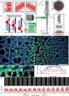
Fig. 1
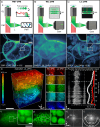
Fig. 1. Concept and resolution improvement of lightsheet line-scanning structured illumination microscopy (LiL-SIM).
a Optical schematic of the LiL-SIM setup. The orientation of the excitation line can be set by the rotation unit, which is composed of a piezoelectric rotation stage, a half-wave plate and a Dove prism. SC galvo-scanner, SL scan lens, TL tube lens, DM dichroic mirror, BFP back focal plane, S sample, F1, F2 filters, L lens. b The emission of neighboring lines is suppressed with the camera’s light sheet shutter mode, leading to less accumulated background compared to rolling shutter mode. c Comparison of two-photon excited fluorescence lightsheet line-scanning microscopy (LiL-2PM) (black), deconvolved LiL-2PM reconstruction (blue), and LiL-SIM reconstruction (red) with 190 nm fluorescent beads. d Fluorescent line pairs (LPs) can be resolved down to a distance of 270 nm in LiL-2PM (black), while LiL-SIM resolves LPs down to 150 nm (red). The data shown in (c) is a representative image out of (N = 5) measurements acquired in distinct bead samples. Scale bars: 5 μm, inset 1 μm. The CAD sketch of the rotation mount in this figure is courtesy of Thorlabs, Inc.
Fig. 2
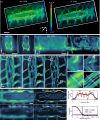
Fig. 2. Comparison of two-photon microscopy modalities and evaluation of modulation contrast for LiL-SIM imaging.
Comparison of signal-to-background and signal-to-noise ratio in two-photon fluorescence microscopy images acquired in Pinus radiata tissue with a photomultiplier tube (PMT), b camera with rolling shutter (RS) mode, and c camera with lightsheet shutter (LSS) mode. The images presented in (a) were acquired with a commercial PMT-based microscope system (PMT-2PM) at the same imaging depth (z = 10 μm) but at distinct lateral positions of the specimen. The signal-to-background ratio is increased when using lightsheet line-scanning two-photon microscopy (LiL-2PM) over widefield line-scanning two-photon microscopy (WiL-2PM). d Normalized two-photon excited raw LiL-2PM volume of zebrafish with penetration depths ranging from 0 to 80 μm. These raw images were all acquired with the same pattern angle and phase at a pattern spacing of 350 nm. e Fourier-transformed planes of the zebrafish stack acquired with LiL-2PM represent the strength of the modulation contrast of the excitation pattern dependent on the imaging depth. The curve visualizes the strength of the modulation peaks for WiL-2PM (red) and LiL-2PM (white). SIM images can be reconstructed with a modulation contrast higher than 0.1. This corresponds to an imaging depth of 56 μm for super-resolution reconstruction. Extracted planes from 40 μm acquired with f WiL-2PM and g LiL-2PM demonstrate the improved modulation contrast achieved with LiL-2PM. h, i Fourier transforms of the images shown in (f, g) visualize the superior modulation contrast achieved with LiL-2PM over WiL-2PM. White rings indicate the spatial frequency in k-space. Data shown in (ac) are representative images taken out of volume stack measurements (N = 5 for each modality). The data and the corresponding contrast enhancement shown in (di) has been verified in (N = 3) zebrafish volume stacks taken at distinct locations. Scale bars: ac 10 μm, insets 3 μm. d 10 μm, inset 5 μm. f, g 10 μm, inset 2 μm.
Fig. 3
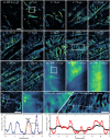
Fig. 3. Extending the penetration depth of super-resolved imaging in Pinus radiata tissue.
a Volume stacks acquired with WiL-2PM and LiL-2PM demonstrate the enhanced optical sectioning effect when using LSS mode. b Axial center cross-sections in xz and yz. c Comparison of WiL-2PM, LiL-2PM and LiL-SIM images of Pinus radiata at a pattern spacing of 300 nm and an imaging depth of 10 μm into the sample. Magnified LiL-SIM insets of the white dashed regions of interest indicate clear resolution improvement compared to the LiL-2PM insets. Line width comparison of LiL-2PM (black) and LiL-SIM reconstruction (red) along the dashed line shown in the insets. FRC data (black) and fit (red) indicate a resolution of 163 nm, taken at a correlation factor of 0.143 (blue bar). d WiL-2PM vs. LiL-SIM at 20 and 30 μm imaging depth. Resolution improvement and contrast enhancement shown in (ac) has been verified in (N = 10) volume stacks acquired at distinct locations. The line profile is a representative curve out of (N = 5) individual measurements. FRC curve was taken out of (N = 5) measurements, evaluated for each presented imaging depth. Scale bars a, b 10 μm. c 5 μm, inset 1 μm. d 2 μm.
Fig. 4
Fig. 4. LiL-SIM imaging in dense mouse heart muscle tissue.
Upper panel: Two-photon excited fluorescence images of mouse heart muscle acquired with LiL-SIM in varying depths from 1 to 70 μm. The depth at which the images were acquired is indicated at the top of each image. Up to an imaging depth of 50 μm, the pattern line spacing was set to 350 nm and to 400 nm for higher imaging depths. The LiL-SIM insets a at 5 μm and d at 55 μm show improved resolution compared to WiL-2PM and LiL-2PM insets (b, c). e Line profile (blue) taken along the dashed line in LiL-SIM inset (a). The distance between Gaussian fit 1 (orange) and Gaussian fit 2 (yellow) is 146 nm, while Gaussian fit 3 (purple) has a FWHM of 177 nm. f Comparison of line profiles from inset (,c, d) between LiL-SIM (red) and LiL-2PM (black). Data shown is a representative volume stack out of (N = 3) stacks taken at distinct locations. Line profiles have been evaluated in (N = 5) individual measurements for each presented imaging depth. Scale bar: 5 μm, left inset 1 μm, right inset 2 μm.

References

    1. Schermelleh, L. et al. Super-resolution microscopy demystified. Nat. Cell Biol.21, 72–84 (2019). - PubMed
    1. Shim, S.-H. et al. Super-resolution fluorescence imaging of organelles in live cells with photoswitchable membrane probes. Proc. Natl. Acad. Sci. USA109, 13978–13983 (2012). - PMC - PubMed
    1. Laine, R. F. et al. Structural analysis of herpes simplex virus by optical super-resolution imaging. Nat. Commun.6, 10.1038/ncomms6980 (2015). - PMC - PubMed
    1. Nizamudeen, Z. et al. Rapid and accurate analysis of stem cell-derived extracellular vesicles with super resolution microscopy and live imaging. Biochim. Biophys. Acta Mol. Cell Res.1865, 1891–1900 (2018). - PMC - PubMed
    1. Möckl, L. & Moerner, W. E. Super-resolution microscopy with single molecules in biology and beyond-essentials, current trends, and future challenges. J. Am. Chem. Soc.142, 17828–17844 (2020). - PMC - PubMed

LinkOut - more resources