. 2025 Dec;17(1):2516676.
doi: 10.1080/19420862.2025.2516676. Epub 2025 Jun 25.

Developing drug-like single-domain antibodies (VHH) from in vitro libraries

Affiliations

Developing drug-like single-domain antibodies (VHH) from in vitro libraries

M Frank Erasmus et al. MAbs. 2025 Dec.

Abstract

Here, we describe a new VHH library for therapeutic discovery which optimizes humanness, stability, affinity, diversity, developability, and facile purification using protein A in the absence of an Fc domain. Four therapeutic humanized VHHs were used as scaffolds, into which we inserted human HCDR1s, HCDR2s and HCDR3s. The HCDR1 and HCDR2 sequences were derived from human VH3 family next-generation sequencing datasets informatically purged of sequence liabilities, synthesized as array-based oligonucleotides, cloned as single CDR libraries into each of the parental scaffolds and filtered for protein A binding by yeast display to ensure correct folding and display. After filtering, the CDR1 and CDR2 libraries were combined with amplified human HCDR3 from human CD19+ IgM+ B cells. This library was further improved by eliminating long consecutive stretches of tyrosines in CDR3 and enriching for CDR1-2 diversity with elevated tolerance to high temperatures. A broad diversity of high affinity (100 pM-10 nM), developable binders was directly isolated, with developability evaluated for most assays using the isolated VHHs, rather than fused to Fc, which is customary. This represents the first systematic developability assessment of isolated VHH molecules.

Keywords: Antibody; VHH; camelid; drug discovery; phage display; single domain; yeast display.


Conflict of interest statement

MFE, EM, LARC, DK, HT, JL, RDN, AF, AD, LS, SD, FF, ARMB are or were employees of Specifica, an IQVIA business at the time of writing. CLL is currently an employee of Sanofi, Cambidge, USA. All other authors declare no conflicts of interest. AART and ARMB are listed as co-inventors on patent describing this technology.

Figures

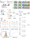
A comparative analysis of VHH frameworks, immunoblot detection, ELISA results, and flow cytometry analysis to evaluate the display and binding capabilities of therapeutic VHHs.
Figure 1.
Scaffolds. (a) Percent identity comparisons between frameworks 1–4 across the four therapeutic VHH, human germline VH3–23, and alpaca germline VH3–3. (b) An immunoblot (anti-SV5) detecting display of the four selected therapeutic VHH on phage particles. The upper band corresponds to the phage pIII linked to the VHH domain, while the lower band represents a degradation product comprising pIII alone. 1-caplacizumab, 2-isecarosmab, 3-sonelokimab, 4-vobarilizumab. (c) Results from a direct ELISA where the four therapeutic phage-VHH were probed against their corresponding immobilized antigens. (d) Flow cytometric analysis of yeast displaying two control scFv, binding (VH3–7) and not binding (VH4–30) SpA, and the four therapeutic VHHs. Yeast cells (blue: induced for display; pink: not induced) were incubated with protein a conjugated to the APC fluorophore. A right shift in fluorescence along the X-axis reflects protein a binding.
Library design schematic, sequence logos displaying human and alpaca CDR diversity, yeast filtering schematic, flow cytometry histograms, NGS sequencing results, CDR3 length distribution, and species accumulation plot to analyze the diversity and functionality of CDRs in VHH libraries.
Figure 2.
CDR diversity. (a) Schematic of the library design highlighting its three primary components: human diversity without sequence liabilities (CDR1–2), natural human diversity sourced from CD19+ IgM+ B-cells (CDR3), and humanized therapeutic VHH scaffold (frameworks 1–3) capable of binding protein a for easy purification. (b) Sequence logos representing natural human VH3 family HCDR diversity (top), human CDRs used to build the library (middle), and natural alpaca (vicugna pacos) VHH diversity (bottom). The height of each letter indicates the frequency of an amino acid at each position. For visual simplicity, only CDR3s with 17 amino acids are shown. (c) A schematic outline demonstrating the yeast filtering method applied to CDR1 and CDR2 in libraries v1 and v2 (heat shock), aimed at selecting CDR functionality and protein a binding in the libraries. (d-g) Histograms show the flow cytometry analysis of yeast cells featuring the four parental VHH (orange), CDR1, and CDR2 libraries prior to protein a filtering (red) and after filtering (blue). The left peaks indicate cells that either do not display the VHH or present molecules that cannot bind protein A. The right peak represents cells displaying a VHH that can bind protein A. (h-k) NGS sequencing results for single CDR libraries before (orange) and after (blue) protein a filtering. (l) Distribution of CDR3 lengths in the library and in the alpaca repertoire are depicted. Therapeutic VHH CDR3 lengths analyzed in the work are indicated with arrows. (m) Species accumulation plot from NovaSeq analysis reveals the diversity of HCDR3 in the cloned library.
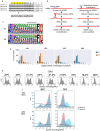
Analysis of poly-tyrosine regions and their targeted degradation, Weblogo representations of HCDR3, NGS analysis for contiguous tyrosines, evaluation of CDR libraries under heat shock, and flow cytometry results for pre- and post-heat shock populations.
Figure 3.
Iterative improvement of the Gen3 VHH design. (a) Poly-tyrosine regions in different human JH6 alleles. The highlighted area was targeted for degradation during cDNA synthesis. (b) A schematic illustration of targeted degradation for poly-tyrosine regions employing specific primers during HCDR3 retrieval from CD19+ cells. After poly-tyrosine encoding mRNAs are degraded by RNAse H, the remaining mRNAs are converted into cDNA using a CH1 primer and HCDR3s amplified from the intact cDNA. (c) Weblogo representations of HCDR3 for v1 (top) and v2, tyrosine depleted, (bottom) libraries. (d) NGS analysis of the naïve library from V1 (blue) and V2 (orange) populations showing the number of contiguous tyrosines. (e) Representative analysis of individual CDR libraries (e.g., CDR1 diversity with constant frameworks alongside other CDRs from library 1) that underwent heat shock and were evaluated across a range of temperatures, identifying an optimal temperature of 76°C. f) analysis from sublibrary 1 of the final cloned output shows pre-heat shock (round 1; red) and post-heat shock (round 5; blue) populations, assessed via flow cytometry for CDR1 (top) and CDR2 (bottom).
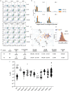
Flow cytometry analysis of VHH-Fc binding to IFNa2, Sanger sequencing results for contiguous tyrosines, isoaffinity plot showing binding affinities, Levenshtein distance analysis of HCDR3 sequences, and summary of binding statistics and affinities for v1 and v2 populations.
Figure 4.
Discovery and affinity characterization of a broad panel of VHH-Fc from V1 and V2 libraries. (a) Flow cytometry analysis of the final sorted population of V1 (top panel) and V2 (bottom panel) binding to varying concentrations of IFNa2 (top to bottom) for each of the different sublibraries (left to right). Display is detected using anti-SV5 conjugated to PE, while binding is detected with biotinylated IFNa2 and streptavidin-alexa-633. (b) Sanger sequencing of clones selected against IFNa2 plotting the number of contiguous tyrosines in the V2 library (orange) compared to V1 (blue). (c) An isoaffinity plot showing on-rate (M-1 s-1) on the y-axis and off-rate (s-1) on the x-axis, with affinities on the diagonal, indicating the wide range of affinities against IFNa2 for VHH-Fc in both v1 (blue) and v2 (orange) populations. (d) Levenshtein distance of the HCDR3 amino acid sequence among the different binders of v1 (blue) and v2 (orange) populations. (e) Summary of binding statistics and affinities for v1 and v2 selected populations. (f) Affinity plots for IFNa2 and ten other undisclosed targets.
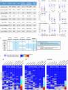
Table describing the library format used and assay threshold, boxplots showing developability metrics of VHH and VHH-Fc, summary of V2 over V1 improvements, and attrition rates across developability assays for different populations.
Figure 5.
Developability of VHH and VHH-Fc from the V1 and V2 libraries. (a) Details of the format (VHH vs. VHH-Fc), assay, and thresholds used to carry out the analysis using 1) GMM, 2) parental range ±2 SD of the mean, or 3) bottom 10% threshold. B-J) boxplot of VHH and VHH-Fc across the different developability assays from using parental range (b) Expression titers (mg/L), (c) Melting temperature (oC), (d) Thermal aggregation at SLS473 (oC), (e) Fraction main peak at A280 with SEC-HPLC, (f) standup monolayer chromatography retention time (min), and polyreactivity (relative absorbance units) with (g) PLE, (h) cardiolipin, (i) BVP, and (j) dsDNA. Boxplots show interquartile range (IQR) from quartile 1 to quartile 3 with the horizonal line showing the median, and whiskers showing the Q1–1.5*IQR and Q3 + 1.5*IQR. Each point represents a unique measurement and is color-coded dark red if worse than the GMM threshold or blue if better than GMM threshold. Horizontal lines represent the GMM threshold (red) or ± 2 SD from parental mean (dark blue). (k) summary of V2 over V1 improvement. If ≥ 5% antibodies show improvement of V2 population over V1 using criteria 1–3 or if there is significant difference of V2 better than V1 (using criteria 4), boxes are scored + 1 and colored light blue. No difference is indicated with 0 and colored white. Total summary of summed scores shown in last column with those ≥ 2 of V2 improvement over V1 colored light blue. L) attrition rate of binders (y-axis) for V1 (left panel), V2 (middle panel), and optimal combination [V1 (lib3)+V2 (lib1,2,4)] across the different developability assays (x-axis).

References

    1. Morrison C. Nanobody approval gives domain antibodies a boost. Nat Rev Drug Discov. 2019;18(7):485–18. doi: 10.1038/d41573-019-00104-w. - DOI - PubMed
    1. Revets H, De Baetselier P, Muyldermans S. Nanobodies as novel agents for cancer therapy. Expert Opin Biol Ther. 2005;5(1):111–124. doi: 10.1517/14712598.5.1.111. - DOI - PubMed
    1. Bannas P, Hambach J, Koch-Nolte F. Nanobodies and nanobody-based human heavy chain antibodies as antitumor therapeutics. Front Immunol. 2017;8:1603. doi: 10.3389/fimmu.2017.01603. - DOI - PMC - PubMed
    1. Hu Y, Liu C, Muyldermans S. Nanobody-based delivery systems for diagnosis and targeted tumor therapy. Front Immunol. 2017;8:1442. doi: 10.3389/fimmu.2017.01442. - DOI - PMC - PubMed
    1. Hamers-Casterman C, Atarhouch T, Muyldermans S, Robinson G, Hammers C, Songa EB, Bendahman N, Hammers R. Naturally occurring antibodies devoid of light chains. Nature. 1993;363(6428):446–448. doi: 10.1038/363446a0. - DOI - PubMed

MeSH terms

Substances

LinkOut - more resources