. 2025 Jun 25;13(1):133.
doi: 10.1186/s40478-025-02048-2.

Copper supplementation mitigates Parkinson-like wild-type SOD1 pathology and nigrostriatal degeneration in a novel mouse model

Affiliations

Copper supplementation mitigates Parkinson-like wild-type SOD1 pathology and nigrostriatal degeneration in a novel mouse model

Benjamin D Rowlands et al. Acta Neuropathol Commun. .

Abstract

Misfolded wild-type superoxide dismutase 1 (disSOD1) protein is implicated in the death of substantia nigra (SN) dopamine neurons in Parkinson disease. Regionally reduced copper availability, and subsequent reduced copper binding to SOD1, is a key factor driving the development of this pathology, suggesting brain copper supplementation may constitute an effective means of preventing its formation. We evaluated whether the blood-brain-barrier-permeable copper delivery drug, CuATSM, attenuated the misfolding and deposition of wild-type disSOD1 and associated neuron death in a novel mouse model that expresses this pathology. These factors were profiled using proteomic and elemental mass spectrometry, together with biochemical and histological workflows. We demonstrated copper supplementation corrects altered post-translational modifications on soluble SOD1 and improves the enzymatic activity of the protein in the brains of these animals. These changes were associated with a significant reduction in disSOD1 pathology and preservation of dopamine neurons in the SN, which were highly correlated with tissue copper levels. Our data position wild-type disSOD1 pathology as a novel drug target for Parkinson disease and suggest that brain copper supplementation may constitute an effective means of slowing SN dopamine neuron death in this disorder.

Keywords: Copper supplementation; CuATSM; Mouse model; Neurodegeneration; Parkinson disease; Post-translational modification; Protein misfolding; Substantia Nigra Pars compacta; Superoxide dismutase 1.


Conflict of interest statement

Declarations. Ethics approval and consent to participate: All experimental procedures involving the use of mice conformed to the Australian Code of Practice for the Care and Use of Animals for Scientific Purposes, with protocols approved by the Animal Ethics Committee at the University of Sydney (Ethics ID: 2020/1849). Consent for publication: All authors read and approved the final manuscript prior to publication. Competing interests: The authors declare no competing interests.

Figures
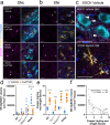
Fig. 1
Fig. 1
Quantification of total disSOD1 burden in the SN of all mouse strains following treatment. Immunofluorescent staining of fixed midbrain tissues from the substantia nigra pars compacta (SNc) and substantia nigra pars reticulata (SNr) of hSOD1WT and SOCK mice treated daily with vehicle or 15 mg/kg CuATSM. Immunostaining utilized the unfolded beta barrel (UβB) conformation-specific SOD1, which revealed disSOD1 aggregates (magenta) within and outside of dopamine neurons (TH, cyan; white arrowheads) and astrocytes (GFAP, yellow; double white arrowheads). Individual panels for immunostaining of the SNc and SNr of SOCK and hSOD1WT mice, as well as Ctr1+/− and wild-type mice, are presented in Supplementary Figs. 13 and 14. Antibody details are presented in Supplementary Table 1. Scale bars represent 20 μm in panels a-c. d. The volume of disSOD1 expressed relative to the total volume of tissue within which it was quantified varied significantly between genotypes and was elevated in SOCK mice compared with hSOD1WT mice treated with vehicle. CuATSM treatment elicited a significant decrease in this pathology in SOCK mice but not in any control mouse strains. e. Midbrain copper levels were decreased in vehicle-treated Ctr1+/− and SOCK mice compared with wild-type mice, while CuATSM treatment induced significant increases in midbrain copper levels in all four mouse strains. Data in panels d and e represent mean ± SEM (n = 10/genotype/treatment), with full details of statistical tests presented in Supplementary Table 2. Comparisons marked with an asterisk (*) denote those made between vehicle- and CuATSM-treated mice of the same genotype, those marked with an arrowhead (^) demarcate those made to vehicle-treated wild-type mice, while those marked with a hashtag (#) denote those made to vehicle-treated hSOD1WT mice. #### p < 0.0001, ****p < 0.0001, ^p < 0.05, ^^p < 0.01. f. DisSOD1 volume in the SN was inversely correlated with midbrain copper content in vehicle- and CuATSM-treated SOCK and hSOD1WT mice. Statistical test details are presented in the panel
Fig. 2
Fig. 2
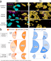
Impact of CuATSM treatment on the distribution of disSOD1 pathology in the SN of SOCK mice. (a) Three-dimensional reconstructions of immunostaining for disSOD1 (UβB), dopamine neurons (TH, cyan) and astrocytes (GFAP, yellow) in the SNc and SNr of SOCK and hSOD1WT mice. DisSOD1 localized within or outside of either cell type is presented in white or magenta, respectively. Scale bars represent 20 μm. Reconstructions of all four mouse strains treated with vehicle or CuATSM are presented in (Supplementary Fig. 15). Panels displayed in this figure constitute those most important for illustrating the higher level of disSOD1 pathology in SOCK mice and the impact of CuATSM treatment. (b) Proportions of disSOD1 pathology colocalized within dopamine (DA) neurons, astrocytes, or other compartments (other). Percentages were generated by scaling the raw volume of disSOD1 in each compartment to the total volume of disSOD1 in the entire SN (SNc + SNr) of vehicle-treated SOCK mice, as this represents the maximum disSOD1 volume exhibited by any mouse strain/treatment combination. Raw disSOD1 volumes are reported in Supplementary Table 3
Fig. 3
Fig. 3
Altered SOD1 PTMs in the SN of vehicle- and CuATSM-treated SOCK mice. Atypical oxidation of SOD1 histidine residues was increased in the SN of SOCK mice compared with hSOD1WT mice (a), while glycosylation (b) and acetylglucosamine (c) were significantly decreased. Side chains of labelled residues are highlighted in black, red and yellow, respectively. Residues are labelled using one letter amino acid codes. Copper and zinc ions are highlighted in orange and cyan respectively. Alterations to SOD1 PTMs in vehicle-treated (d) and CuATSM-treated (e) SOCK mice. Circles for each type of modification in the legend are scaled to the number of residues where that PTM is altered in each treatment group. GlyGly modifications result from tryptic digestion of ubiquitin-conjugated proteins, which serve as indicators of protein ubiquitination. Complete details of statistical analyses identifying PTM alterations in vehicle- and CuATSM-treated SOCK mice are presented in Supplementary Table 4. Abbreviations: GlcNAc, acetylglucosamination
Fig. 4
Fig. 4
SOD1 enzymatic activity and protein expression in the SN of all mouse strains following treatment. (a) Total SOD activity varied significantly between mouse strains and was increased in vehicle-treated SOCK and hSOD1WT mice compared with wild-type (WT) and Ctr1+/− mice, as well as vehicle-treated SOCK mice compared with hSOD1WT mice. CuATSM treatment elicited increases in total SOD activity across all strains. (b) SOD activity per unit of SOD1 protein varied significantly between mouse strains and was decreased in vehicle-treated SOCK and hSOD1WT mice compared with wild-type mice, as well as vehicle-treated SOCK mice compared with hSOD1WT mice. CuATSM treatment increased SOD activity per unit of SOD1 protein in SOCK and hSOD1WT mice. (c) SOD activity per unit of SOD1 protein was positively correlated with midbrain copper content in vehicle- and CuATSM-treated SOCK and hSOD1WT mice. (d) DisSOD1 volume in the SN was inversely correlated with SOD activity per unit of SOD1 protein in vehicle- and CuATSM-treated SOCK mice. (e) SOD1 protein levels varied significantly between mouse strains and were increased in vehicle-treated SOCK and hSOD1WT mice compared with wild-type and Ctr1+/− mice. CuATSM treatment elicited increases in SOD1 protein levels in wild-type and Ctr1+/− mice. Full representative immunoblots are presented in Supplementary Fig. 18. Data in panels a, b and e represent mean ± SEM (n = 9–11/genotype/treatment), with full details of statistical tests presented in Supplementary Table 2. Comparisons marked with an asterisk (*) denote those made between vehicle- and CuATSM-treated mice of the same genotype, those marked with an arrowhead (^) demarcate those made to vehicle-treated wild-type mice, while those marked with a hashtag (#) denote those made to vehicle-treated hSOD1WT mice. ## p < 0.01, ### p < 0.001, *p < 0.05, ****p < 0.0001, ^^^^p < 0.0001. (f) SOD1 protein levels were positively correlated with total SOD activity in vehicle- and CuATSM-treated wild-type and Ctr1+/− mice. Statistical test details for c, d and f are presented in the panel
Fig. 5
Fig. 5
Evaluation of nigrostriatal neurotransmission and neurodegeneration in all mouse strains following treatment. (a) Striatal dopamine levels were significantly reduced in hSOD1WT and SOCK mice treated with vehicle compared with wild-type mice, whilst CuATSM treatment increased striatal dopamine levels in SOCK mice and decreased them in wild-type mice. (b) Striatal dopamine turnover, defined as the levels of homovanillic acid (HVA) and DOPAC normalized to dopamine (DA) levels, was significantly elevated in SOCK mice treated with vehicle compared with wild-type mice, with CuATSM treatment preventing this alteration and eliciting an elevation in dopamine turnover in wild-type and hSOD1WT mice. (c) Stereological quantification of 3D-reconstructed tyrosine hydroxylase immunostaining revealed a significant loss of SNc dopamine neurons in vehicle-treated SOCK mice compared with all control mouse strains, which was rescued by CuATSM treatment. Representative 3D reconstructions of each genotype/treatment are presented in Supplementary Fig. 15. Data in panels a-c represent mean ± SEM (n = 9–11/genotype/treatment), with full details of statistical tests presented in Supplementary Table 2. Comparisons marked with an asterisk (*) denote those made between vehicle- and CuATSM-treated mice of the same genotype, those marked with an arrowhead (^) demarcate those made to vehicle-treated wild-type mice. *p < 0.05, ***p < 0.001, ^p < 0.05, ^^p < 0.01, ^^^p < 0.001. The density of SNc dopamine neurons was positively correlated with midbrain copper levels (d), inversely correlated with SNc disSOD1 volume (e) and positively correlated with midbrain total SOD activity (f) in SOCK mice. Statistical test details for d - f are presented in the panel
Fig. 6
Fig. 6
Motor performance testing in all mouse strains following treatment. In balance beam testing, the latency in time taken to cross the beam (a), together with the number of paw slips (b), varied significantly between mouse strains, with both increased in vehicle-treated SOCK mice compared with wild-type (WT) and hSOD1WT mice. CuATSM treatment significantly reduced both measures in SOCK mice, but did not change them in other strains. By contrast there was no variation in grip strength (c) or open field test performance metrics - time (sec) immobile (d), total distance travelled (meters; e), central zone entries (f)– between all strains and treatment groups. g. Mouse body weight increased at comparable rates between all four mouse strains treated with vehicle (p = 0.9093, F = 0.3890), although CuATSM treatment improved weight gain in SOCK mice (statistical test details presented in panel). h. Spinal motor neuron densities in each mouse genotype did not vary between vehicle and CuATSM treatment, nor did they vary between genotypes within the same treatment group. Data in panels a-f and h represent mean ± SEM (n = 9–11/genotype/treatment), with full details of statistical tests presented in Supplementary Table 2. Comparisons marked with an asterisk (*) denote those made between vehicle- and CuATSM-treated mice of the same genotype, those marked with an arrowhead (^) demarcate those made to vehicle-treated wild-type mice, while those marked with a hashtag (#) denote those made to vehicle-treated hSOD1WT mice. #### p < 0.0001, ****p < 0.0001, ^^^^p < 0.0001

References

    1. Feigin VL et al (2017) Global, regional, and National burden of neurological disorders during 1990–2015: a systematic analysis for the global burden of disease study 2015. Lancet Neurol 16:877–897 - PMC - PubMed
    1. Dorsey ER, Collaborators GPsD (2018) Global, regional, and National burden of parkinson’s disease, 1990–2016: a systematic analysis for the global burden of disease study 2016. Lancet Neurol 17:939–953 - PMC - PubMed
    1. Dorsey ER, Sherer T, Okun MS, Bloem BR (2018) The emerging evidence of the Parkinson pandemic. J Parkinsons Dis 8:S3–S8 - PMC - PubMed
    1. Bloem BR, Okun MS, Klein C (2021) Parkinson’s disease. Lancet 397:2284–2303 - PubMed
    1. Höglinger GU et al (2024) A biological classification of parkinson’s disease: the synneurge research diagnostic criteria. Lancet Neurol 23:191–204 - PubMed

Publication types

MeSH terms

LinkOut - more resources