. 2025 Jul;34(7):e70205.
doi: 10.1002/pro.70205.

Prokaryotic mechanosensitive channels mediate copper influx

Affiliations

Prokaryotic mechanosensitive channels mediate copper influx

Yara Ghnamah et al. Protein Sci. 2025 Jul.

Abstract

Copper is an essential micronutrient in all kingdoms of life, requiring a meticulous balance between acquisition and toxic overload. While copper import in eukaryotes has been investigated extensively, few prokaryotic copper importers have been identified, leading to the notion that cytoplasmic copper uptake is unnecessary in prokaryotes. Here we report that mechanosensitive channels are key players in prokaryotic copper import. Deletion of the gene encoding the Escherichia coli small mechanosensitive channel, EcMscS, leads to significantly reduced copper influx. Conversely, overexpression of EcMscS leads to increased copper influx, elevated intracellular copper content, and renders cells hypersensitive to copper. Furthermore, specific channel blockers and competing permeating ions inhibit EcMscS copper conductance, lowering intracellular copper accumulation and alleviating copper hypersensitivity. These findings extend beyond E. coli, as other prokaryotic small mechanosensitive channels of bacterial and archaeal origin also facilitate copper influx. Taken together, these results uncover a previously unknown moonlighting function for mechanosensitive channels as a pathway for prokaryotic copper uptake.

Keywords: bacteria; copper; ion channels; mechanosensitive channels; membrane permeation; metal homeostasis; prokaryotes; transport.


Conflict of interest statement

The authors declare no conflicting interests.

Figures

FIGURE 1
FIGURE 1
ECMscS facilitates copper permeation. (a) At the time indicated by the arrow, 100 μM CuSO4 was injected to cultures of WT or ΔmscS cells (red and blue curves, respectively) expressing the copper reporter pcopA‐Lux. Luminescence was normalized by the optical density of the cultures. (b) Same as A, only the cells were pre‐exposed to isosmotic or mild hypoosmotic conditions (open and closed circles, respectively. See methods for full details). Results are the mean of biological triplicates, with error bars indicating standard deviations.
FIGURE 2
FIGURE 2
ECMscS overexpression increases copper sensitivity. (a) Cells transformed with an empty control vector or the ECMscS expression vector pMscS (blue and red curves, respectively) were cultured in the absence or presence of 2.5 mM CuSO4 (open and closed symbols, respectively). Optical density at 600 nm was measured every 5 min. (b) Cells transformed with an empty control vector or the ECMscS expression vector pMscS (blue and red curves, respectively) were cultured for 10 h in the presence of the indicated concentration of CuSO4 in the absence or presence of 0.01 mM IPTG (open and closed symbols, respectively). Growth in the absence of cooper was defined as 100%. Averages of biological triplicates are shown, with error bars indicating standard deviations (shown unless smaller than icons).
FIGURE 3
FIGURE 3
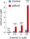
ECMscS expression increases intracellular copper accumulation. (a) Cells transformed with an empty control vector or the ECMscS expression vector pMscS (blue and red bars, respectively) were cultured in the presence of IPTG (0.01 mM) to mid log phase and then exposed for 30 min to the indicated concentrations of CuSO4. Cells were washed with an EDTA containing buffer and their intracellular copper concentration was determined by ICP‐MS. Shown are averages of biological triplicates, with error bars indicating standard deviations. ***p < 0.005; ns, not significant, as determined by a two‐tailed Student's t‐test.
FIGURE 4
FIGURE 4
Copper sensitivity correlates with the activity level of ECMscS. Cells transformed with an empty control vector or vectors encoding EcMscS WT or A106V (blue, red, and green curves, respectively, as indicated) were cultured in the absence (a) or presence (b) of 2.5 mM CuSO4. (c) Cells transformed with a vector encoding WT ECMscS (all curves) were cultured in the absence or presence of 2.5 mM CuSO4 (black and red curves, respectively) and in the absence or presence of 2 μM GsMTx4 inhibitor (open and closed symbols, respectively). The dashed line represents data points used to generate the bar graph shown in D. (d) Optical density (OD600) at 800 min (dashed vertical line in C) of cells grown in the absence or presence of 2.5 mM CuSO4 and in the absence (−) or presence (+) of 2 μM GsMTx4 inhibitor, as indicated. Results are averages of biological triplicates with error bars indicating standard deviations (shown unless smaller than the icons). **p < 0.02; ns, not significant, as determined by a two‐tailed Student's t‐test.
FIGURE 5
FIGURE 5
Competing ions reduce EcMscS‐mediated copper influx. Cells transformed with the ECMscS expression vector pMscS were cultured in LB media in the presence or absence of copper (2.5 mM), lithium (12.5 mM), and calcium (5 mM), as indicated. The optical density at 600 nm was measured after 10 h of growth. (B) Copper content of cells expressing EcMscS exposed for 30 min to the indicated metals (or their combinations). Averages of biological triplicates are shown, with error bars indicating standard errors (shown unless smaller than icons). ***p < 0.001; ns, not significant, as determined by a one‐way ANOVA and Tukey HSD.
FIGURE 6
FIGURE 6
EcMscS mediates influx of additional metals. (a) Cells transformed with an empty control vector or the ECMscS expression vector pMscS (blue and red curves, respectively) were cultured in the presence of 250 μM CdCl2. Optical density at 600 nm was measured every 5 min. (b) Cells transformed with an empty control vector or the ECMscS expression vector pMscS (blue and red curves, respectively) were cultured in the presence of the indicated concentrations of ZnSO4, and the optical density at 600 nm was measured after 10 h of growth. Shown is the growth, in percent, relative to the growth in the absence of metal. (c) Metal content of control or EcMscS‐expressing cells exposed for 30 min to either 250 μM CdCl2 or 1.25 mM ZnSO4, as indicated. ***p < 0.001; **p < 0.005; as determined by a two‐tailed Student's t‐test. In (a)–(c), results are averages of biological triplicates, with error bars representing standard errors (shown unless smaller than the icons).
FIGURE 7
FIGURE 7
Copper conductance is conserved among prokaryotic mechanosensitive channels. (a) Cells were transformed with an empty control vector or vectors encoding MscS homologues from the indicated organisms, and their intracellular content was determined using ICP‐MS following exposure to 2.5 mM CuSO4. (b) Cells transformed with an empty control vector (blue curve) or vectors encoding MscS homologues from Thermus thermophilus, Thermotoga maritima, or Salmonella enterica (green, orange, and red curves, respectively) were cultured in the presence of the indicated concentrations of CuSO4, and the optical density at 600 nm was measured after 10 h of growth. Averages of biological triplicates are shown, with some error bars (indicating standard deviations) not visible as they are smaller than icons.

Update of

References

    1. Bae C, Sachs F, Gottlieb PA. The mechanosensitive ion channel Piezo1 is inhibited by the peptide GsMTx4. Biochemistry. 2011;50(29):6295–6300. 10.1021/bi200770q - DOI - PMC - PubMed
    1. Bandara T, Xu J, Potter ID, Franks A, Chathurika JBAJ, Tang C. Mechanisms for the removal of Cd(II) and Cu(II) from aqueous solution and mine water by biochars derived from agricultural wastes. Chemosphere. 2020;254:126745. 10.1016/j.chemosphere.2020.126745 - DOI - PubMed
    1. Bass RB, Strop P, Barclay M, Rees DC. Crystal structure of Escherichia coli MscS, a voltage‐modulated and mechanosensitive channel. Science. 2002;298(5598):1582–1587. 10.1126/science.1077945 - DOI - PubMed
    1. Booth IR, Edwards MD, Black S, Schumann U, Miller S. Mechanosensitive channels in bacteria: signs of closure? Nat Rev Microbiol. 2007;5(6):431–440. 10.1038/nrmicro1659 - DOI - PubMed
    1. Booth IR, Miller S, Müller A, Lehtovirta‐Morley L. The evolution of bacterial mechanosensitive channels. Cell Calcium. 2015;57(3):140–150. 10.1016/j.ceca.2014.12.011 - DOI - PubMed

MeSH terms