. 2025 May 23;14(6):624.
doi: 10.3390/antiox14060624.

Antioxidant Effect of a Fucus vesiculosus Extract on Intestinal Ischemia/Reperfusion Injury in Rats: A Biochemical and Histological Study

Affiliations

Antioxidant Effect of a Fucus vesiculosus Extract on Intestinal Ischemia/Reperfusion Injury in Rats: A Biochemical and Histological Study

Desirée Sánchez-Bonet et al. Antioxidants (Basel). .

Abstract

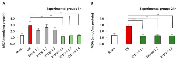
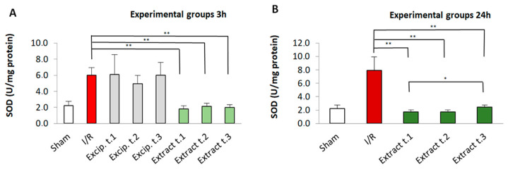
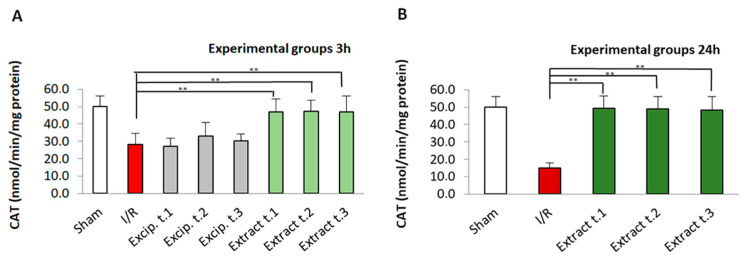
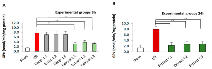
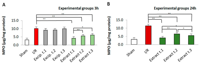
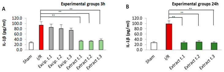
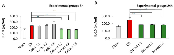
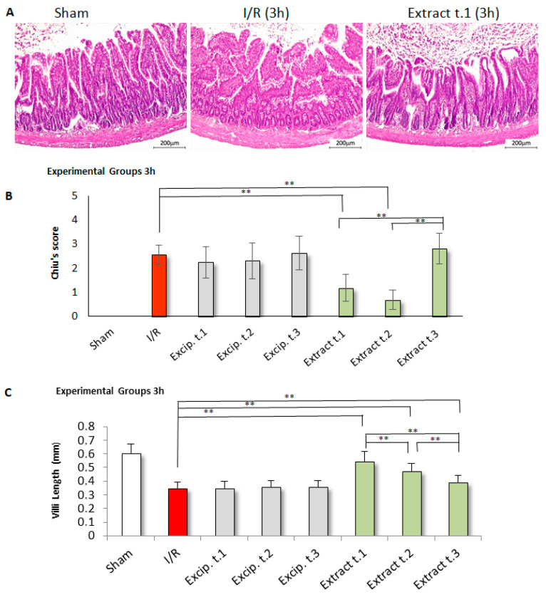
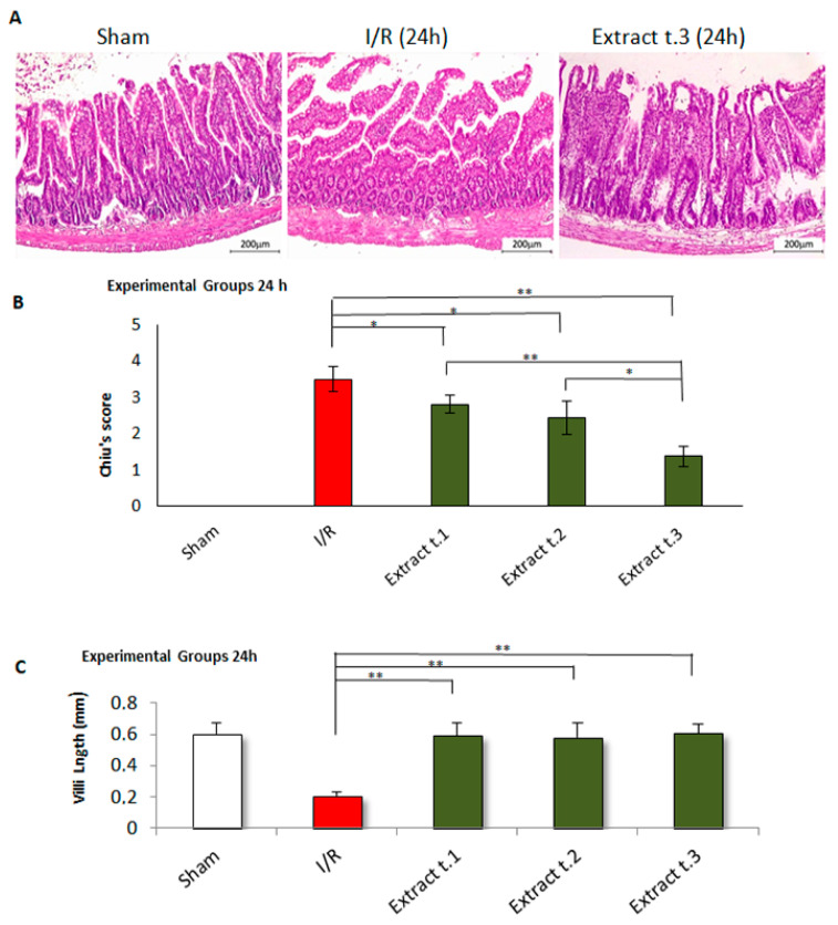
Fucus vesiculosus is a brown seaweed known for its strong antioxidant properties, mainly attributed to its high polyphenolic content. This study aimed to evaluate the antioxidant protective effect of an optimised F. vesiculosus extract in an experimental model of intestinal ischemia/reperfusion (I/R) injury, considering the intestine as particularly vulnerable to this pathology. Seventy-two male Wistar albino rats were randomly divided into twelve groups: Sham, I/R groups (3 and 24 h reperfusion), I/R plus vehicle groups (three application times, 3 h reperfusion), and I/R plus F. vesiculosus extract groups (three application times, 3 and 24 h reperfusion). Intestinal injury was assessed through biochemical markers (malondialdehyde [MDA], superoxide dismutase [SOD], catalase [CAT], glutathione peroxidase [GPx], and mieloperoxidase [MPO]), inflammatory cytokines (interleukin 1 β [IL-1β] and interleukin [IL-10]), and histological analysis. Results demonstrated that treatment with F. vesiculosus significantly reduced oxidative stress and inflammation caused by I/R injury (p < 0.05), restoring analysed parameters (MDA, SOD, CAT, IL-10) to levels comparable to the Sham group. Histological examination confirmed the preservation of intestinal mucosal integrity following F. vesiculosus administration. These findings suggest that the antioxidant extract from F. vesiculosus effectively protects against intestinal I/R injury, highlighting its potential for clinical use in preventing and managing this pathological condition, particularly in surgical contexts.

Keywords: Fucus vesiculosus; antioxidant; intestinal ischemia/reperfusion; polyphenols.


Conflict of interest statement

The authors declare no conflicts of interest.

Figures

Figure 1
Figure 1
MDA levels present in the different experimental groups: (A) 60 min of ischemia and 3 h of reperfusion; (B) 60 min of ischemia and 24 h of reperfusion. Values expressed in nmol/mg protein (mean ± standard deviation). Significant differences between the treatment groups and the I/R group are indicated (** p < 0.01); Mann–Whitney U test, SPSS.
Figure 2
Figure 2
SOD levels present in the different experimental groups: (A) 60 min of ischemia and 3 h of reperfusion; (B) 60 min of ischemia and 24 h of reperfusion). Values expressed in U/mg protein (mean ± standard deviation. Significant differences between the treatment groups and the I/R group are indicated (* p < 0.05, ** p < 0.01); Mann–Whitney U test, SPSS.
Figure 3
Figure 3
CAT levels present in the different experimental groups: (A) 60 min of ischemia and 3 h of reperfusion; (B) 60 min of ischemia and 24 h of reperfusion. Values expressed in nmol/min/mg protein (mean ± standard deviation). Significant differences between the treatment groups and the I/R group are indicated (** p < 0.01); Mann–Whitney U test, SPSS.
Figure 4
Figure 4
GPx levels present in the different experimental groups: (A) 60 min of ischemia and 3 h of reperfusion; (B) 60 min of ischemia and 24 h of reperfusion. Values expressed in nmol/min/mg protein (mean ± standard deviation. Significant differences between the treatment groups and the I/R group are indicated (** p < 0.01); Mann–Whitney U test, SPSS.
Figure 5
Figure 5
MPO levels present in the different experimental groups: (A) 60 min of ischemia and 3 h of reperfusion; (B) 60 min of ischemia and 24 h of reperfusion. Values expressed in µg/mg protein (mean ± standard deviation). Significant differences between the treatment groups and the I/R group are indicated (* p < 0.05, ** p < 0.01); Mann–Whitney U test, SPSS.
Figure 6
Figure 6
Il-1β levels present in the different experimental groups: (A) 60 min of ischemia and 3 h of reperfusion; (B) 60 min of ischemia and 24 h of reperfusion. Values expressed in µg/mg protein (mean ± standard deviation). Significant differences between the treatment groups and the I/R group are indicated (** p < 0.01); Mann–Whitney U test, SPSS.
Figure 7
Figure 7
Il-10 levels present in the different experimental groups: (A) 60 min of ischemia and 3 h of reperfusion; (B) 60 min of ischemia and 24 h of reperfusion. Values expressed in µg/mg protein (mean ± standard deviation. Significant differences between the treatment groups and the I/R group are indicated (** p < 0.01); Mann–Whitney U test, SPSS.
Figure 8
Figure 8
Histological results for the experimental groups after 3 h of reperfusion: (A) Tissue sections of small intestinal mucosa stained with H&E (100× magnification) revealed significant villus fragmentation and loss in the I/R group. In contrast, the villous structure was better preserved in the F. vesiculosus extract group. (B) Distribution of intestinal injury according to the Chiu scoring system (G0 = normal mucosa; G1 = development of subepithelial space at the villus tip, often accompanied by oedema and vascular congestion; G2 = detachment of the epithelial layer from the lamina propria with moderate extension of the subepithelial space, villus tip fragmentation, and haemorrhage; G3 = partial loss of villus tips, extensive epithelial detachment, and fragmentation with loss of the upper third of the villi; G4 = dilated and exposed capillaries with lost villi, though crypts remain present; G5 = haemorrhage, ulceration, disintegration of the lamina propria, and complete mucosal necrosis) indicated that the I/R group exhibited significant alterations, including fragmentation of the upper villus regions and loss of crypts (C) Furthermore, a marked reduction in villus length was noted in the I/R group compared to F. vesiculosus extracts with significant differences (** p < 0.01).
Figure 9
Figure 9
Histological results for the experimental groups after 24 h of reperfusion: (A) Tissue sections of small intestinal mucosa stained with H&E (100× magnification) revealed significant villus fragmentation and loss in the I/R group. In contrast, the villous structure was better preserved in the F. vesiculosus extract group. (B) Distribution of intestinal injury according to the Chiu scoring system (G0 = normal mucosa; G1 = development of subepithelial space at the villus tip, often accompanied by oedema and vascular congestion; G2 = detachment of the epithelial layer from the lamina propria with moderate extension of the subepithelial space, villus tip fragmentation, and haemorrhage; G3 = partial loss of villus tips, extensive epithelial detachment, and fragmentation with loss of the upper third of the villi; G4 = dilated and exposed capillaries with lost villi, though crypts remain present; G5 = haemorrhage, ulceration, disintegration of the lamina propria, and complete mucosal necrosis) indicated that the I/R group exhibited significant alterations, including fragmentation of the upper villus regions and loss of crypts (C) Furthermore, a marked reduction in villus length was noted in the I/R group compared to F. vesiculosus extracts with significant differences (* p < 0.05, ** p < 0.01).

Similar articles

Cited by

References

    1. Aranda-Rivera A.K., Cruz-Gregorio A., Arancibia-Hernández Y.L., Hernández-Cruz E.Y., Pedraza-Chaverri J. RONS and oxidative stress: An overview of basic concepts. Oxygen. 2022;2:437–478. doi: 10.3390/oxygen2040030. - DOI
    1. Kotha R.R., Tareq F.S., Yildiz E., Luthria D.L. Oxidative stress and antioxidants—A critical review on in vitro antioxidant assays. Antioxidants. 2022;11:2388. doi: 10.3390/antiox11122388. - DOI - PMC - PubMed
    1. Namirah I., Wimbanu K.S., Rompies A.M.E., Prayogo Y.S., Arozal W., Fadilah, Hanafi M., Hardiany N.S. The effect of ethanol-based coriander (Coriandrum sativum L.) seed extract on oxidative stress, antioxidant level and cellular senescence in the heart of obese rat. J. Pharm. Pharmacogn. Res. 2024;12:1111–1120. doi: 10.56499/jppres24.1957_12.6.1111. - DOI
    1. Di Meo S., Napolitano G., Venditti P. Physiological and pathological role of ROS: Benefits and limitations of antioxidant treatment. Int. J. Mol. Sci. 2019;20:4810. doi: 10.3390/ijms20194810. - DOI - PMC - PubMed
    1. Sies H. Oxidative stress. Acad. Press. 1985;86:715–748. doi: 10.1146/annurev-biochem-061516-045037. - DOI - PubMed

LinkOut - more resources