. 2025 Jun 9;14(6):700.
doi: 10.3390/antiox14060700.

CircRNA_1156 Attenuates Neodymium Nitrate-Induced Hepatocyte Ferroptosis by Inhibiting the ACSL4/PKCβII Signaling Pathway

Affiliations

CircRNA_1156 Attenuates Neodymium Nitrate-Induced Hepatocyte Ferroptosis by Inhibiting the ACSL4/PKCβII Signaling Pathway

Ning Wang et al. Antioxidants (Basel). .

Abstract

Ferroptosis, a form of regulated cell death driven by lipid peroxidation, has been implicated in the pathogenesis of liver diseases. This study investigates the role of circRNA_1156 in neodymium nitrate (Nd(NO3)3)-induced hepatocyte ferroptosis. Our in vitro experiments revealed that exposure to Nd(NO3)3 (1.2 µM) significantly reduced the viability of AML12 hepatocytes (p < 0.01), increased levels of reactive oxygen species (ROS) and malondialdehyde (MDA) (p < 0.001), and depleted glutathione (GSH) (p < 0.001). However, overexpression of circRNA_1156 effectively reversed these effects and suppressed the expression of ACSL4 and PKCβII (p < 0.01). In our in vivo experiments, chronic exposure to Nd(NO3)3 (7-55 mg/kg for 180 days) induced hepatic iron deposition, mitochondrial damage, and activation of the ACSL4/PKCβII pathway (p < 0.01). These adverse effects were significantly ameliorated by circRNA_1156 overexpression (p < 0.05). Our findings identify circRNA_1156 as a novel inhibitor of Nd(NO3)3-induced ferroptosis via downregulation of the ACSL4/PKCβII pathway, providing valuable therapeutic insights for hepatotoxicity caused by rare earth element compounds.

Keywords: ACSL4/PKCβII pathway; circRNA_1156; ferroptosis; hepatocyte injury; neodymium nitrate.


Conflict of interest statement

The authors declare that this study was conducted in the absence of any commercial or financial relationships that could be construed as potential conflicts of interest.

Figures

Figure 1
Figure 1
Effect of Nd(NO3)3 on the viability of AML12 cells. Cell viability was assessed using the CCK-8 assay after 24 h of exposure to Nd(NO3)3 (1.2 µM). Data are presented as the mean ± SE (n = 3 independent experiments). ** p < 0.01, *** p < 0.001, **** p < 0.0001 compared to the control group.
Figure 2
Figure 2
Detection of intracellular ROS, mitochondrial ROS, and ferroptosis-related markers in AML12 cells after 24-h exposure to 1.2 µM Nd(NO3)3. (A) Detection of intracellular ROS using the DCFH-DA probe. (B) Quantitative analysis of intracellular ROS. (C) Quantitative analysis of mitochondrial ROS in control and circRNA_1156 overexpression cells. (D) Quantitative analysis of mitochondrial ROS. (E) Measurement of mitochondrial membrane potential (Rhodamine 123). (F) Determination of superoxide dismutase (SOD) levels. (G) Determination of malondialdehyde (MDA) levels. (H) Determination of glutathione (GSH) levels. Data are presented as the mean ± SE (n = 3 independent experiments). ** p < 0.01, *** p < 0.001, **** p < 0.0001 compared to the control group.
Figure 3
Figure 3
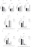
The effect of Nd(NO3)3 on ferroptosis-related gene expression in AML12 cells. (A) Gene expression of circRNA_1156. (B) Gene expression of ACSL4. (C) Gene expression of LPCAT3. (D) Gene expression of PKCβII. (E) Gene expression of circRNA_1156 after transfection with circRNA_1156 overexpression plasmid. (F) Gene expression of ACSL4 after transfection with circRNA_1156 overexpression plasmid. (G) Gene expression of LPCAT3 after transfection with circRNA_1156 overexpression plasmid. (H) Gene expression of PKCβII after transfection with circRNA_1156 overexpression plasmid. Legend: “−/−”: No circRNA_1156 overexpression plasmid transfection and no 24-h exposure to 1.2 µM Nd(NO3)3 (double negative control); “−/+”: No circRNA_1156 overexpression plasmid transfection, but with 24-h exposure to 1.2 µM Nd(NO3)3 (single treatment control); “+/−”: circRNA_1156 overexpression plasmid transfection without 24-h exposure to 1.2 µM Nd(NO3)3 (single genetic manipulation group); “+/+”: circRNA_1156 overexpression plasmid transfection plus 24-h exposure to 1.2 µM Nd(NO3)3 (full treatment group). Data are presented as the mean ± SE (n = 3 independent experiments). * p < 0.05, ** p < 0.01, *** p < 0.001, **** p < 0.0001 compared to the control group.
Figure 4
Figure 4
Effects of Nd(NO3)3 on the expression of ferroptosis-related proteins in AML12 cells. (A) Expression of ferroptosis-related proteins in AML12 cells treated with Nd(NO3)3. (B) Gene expression of circRNA_1156 after transfection with circRNA_1156 overexpression plasmid. (C) Expression of ferroptosis-related proteins in AML12 cells after transfection with circRNA_1156 overexpression plasmid. (D) Grayscale analysis of ACSL4 protein. (E) Grayscale analysis of LPCAT1 protein. (F) Grayscale analysis of PKCβII protein. (G) Grayscale analysis of GPX4 protein. (H) Grayscale analysis of SLC7A11 protein. Legend: “−/−”: No circRNA_1156 overexpression plasmid transfection and no 24-h exposure to 1.2 µM Nd(NO3)3 (double negative control); “−/+”: No circRNA_1156 overexpression plasmid transfection, but with 24-h exposure to 1.2 µM Nd(NO3)3 (single treatment control); “+/−”: circRNA_1156 overexpression plasmid transfection without 24-h exposure to 1.2 µM Nd(NO3)3 (single genetic manipulation group); “+/+”: circRNA_1156 overexpression plasmid transfection plus 24-h exposure to 1.2 µM Nd(NO3)3 (full treatment group). Data are presented as the mean ± SE (n = 3 independent experiments). * p < 0.05, **** p < 0.0001 compared to the control group.
Figure 5
Figure 5
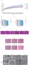
Effects of Nd(NO3)3 on body weight, liver weight, organ index, and pathological histology in C57BL/6J mice. (A) Changes in body weight over 180 days. (B) Absolute liver weight. (C) Liver-to-body weight ratio. (D) Hematoxylin and Eosin (H&E) staining of liver tissues. Scale bar = 100 µm. (E) Prussian blue staining for iron deposition in liver tissues. Magnified areas are indicated with arrows, and iron deposits are annotated. Scale bar = 50 µm. (F) Transmission electron microscopy of liver tissues. Legend: Liver tissues were stained with H&E to visualize cellular structures and with Prussian blue to detect iron deposits. Magnified areas highlight the presence of iron deposits, which appear as blue-black granules. Scale bars are included to provide a reference for the size of the structures shown. Data are presented as the mean ± SE (n = 6 independent experiments).
Figure 6
Figure 6
Effects of Nd(NO3)3 on fluorescence in situ hybridization, immunofluorescence, and ferroptosis biomarkers in C57BL/6J mice. (A) Fluorescence in situ hybridization (FISH) for circRNA_1156. (B) Immunofluorescence staining for GPX4. (C) Immunofluorescence staining for ACSL4. (D) Immunofluorescence staining for LPCAT1. (E) Immunofluorescence staining for PKCβII. (F) SOD activity levels. (G) MDA levels. (H) GSH levels. Data are presented as the mean ± SE (n = 3 independent experiments). * p < 0.05, ** p < 0.01, *** p < 0.001, **** p < 0.0001 compared to the control group.
Figure 7
Figure 7
Molecular pathway diagram showing circRNA_1156 targeting and regulating the expression of ACSL4 and PKCβII to block ferroptosis. Legends: Color Definitions: Red arrows: Indicate the Nd(NO3)3-induced mitochondrial damage pathway (ROS↑ → MDA↑ → GSH↓ → ΔΨm↓); Green arrows: Represent the rescue effects of circRNA_1156 overexpression (ROS↓ → MDA↓ → GSH↑ → restored cell viability); Black arrows: Denote increases (↑) or decreases (↓) in specific indicators.

References

    1. Hu X.F., Jiang J. Ferroptosis and cancer: Interlinkages and potential applications. Clin. Transl. Rep. 2023;1:12–20. doi: 10.58832/ctr.2023.7.6.2. - DOI
    1. Fang X.L., Ding S.Y., Du X.Z., Wang J.H., Li X.L. Ferroptosis—A Novel Mechanism with Multifaceted Actions on Stroke. Front. Neurol. 2022;13:881809. doi: 10.3389/fneur.2022.881809. - DOI - PMC - PubMed
    1. Ding K.Y., Liu C.B., Li L., Yang M., Jiang N., Luo S.L., Sun L. Acyl-CoA synthase ACSL4: An essential target in ferroptosis and fatty acid metabolism. Chin. Med. J. 2023;136:2521. doi: 10.1097/CM9.0000000000002533. - DOI - PMC - PubMed
    1. Wang Y., Yan D., Liu J.B., Tang D.L., Chen X. Protein modification and degradation in ferroptosis. Redox Biol. 2024;75:103259. doi: 10.1016/j.redox.2024.103259. - DOI - PMC - PubMed
    1. Wang Y.X., Xia S.Y. Relationship Between ACSL4-Mediated Ferroptosis and Chronic Obstructive Pulmonary Disease. Int. J. Chronic Obstr. Pulm. Dis. 2023;18:99–111. doi: 10.2147/COPD.S391129. - DOI - PMC - PubMed

LinkOut - more resources