. 2025 Jun 11;14(6):709.
doi: 10.3390/antiox14060709.

Targeting Spermine Oxidase to Mitigate Traumatic Brain Injury Pathology in the Aging Brain

Affiliations

Targeting Spermine Oxidase to Mitigate Traumatic Brain Injury Pathology in the Aging Brain

Jui-Ming Sun et al. Antioxidants (Basel). .

Abstract

Traumatic brain injury (TBI) in the elderly is frequently associated with worsened neurological outcomes and prolonged recovery, yet the age-specific molecular mechanisms driving this vulnerability remain poorly understood. Aging is characterized by increased oxidative stress and chronic neuro-inflammation, both of which may amplify the brain's susceptibility to injury. In this study, we identify spermine oxidase (SMOX), a polyamine-catabolizing enzyme that produces reactive oxygen species, as a key mediator linking oxidative stress and neuro-inflammation to age-dependent TBI susceptibility. Using a mouse model of controlled cortical impact (CCI), we found that SMOX expression was significantly upregulated in aged brains, primarily in neurons and microglia, and this increase correlated with greater microglial activation, elevated pro-inflammatory cytokine expression, and widespread neuronal degeneration. Notably, SMOX upregulation also impaired astrocytic glutamate clearance by disrupting the membrane localization of the transporter GLT-1, contributing to excitotoxic stress. Importantly, analysis of postmortem human brain samples and transcriptomic data revealed a parallel age-related increase in SMOX expression, supporting its translational relevance. The pharmacological inhibition of SMOX with JNJ-9350 in aged mice reduced oxidative and inflammatory markers, preserved neuronal viability, and improved motor, cognitive, and emotional outcomes up to 30 days post-injury. These findings establish SMOX as a critical molecular driver of age-related vulnerability to TBI and highlight its inhibition as a promising therapeutic strategy for improving outcomes in elderly TBI patients.

Keywords: aging brain; brain injury therapeutics; neuro-inflammation; neuronal cell death; oxidative stress; spermine oxidase; traumatic brain injury.


Conflict of interest statement

The authors declare no conflicts of interest.

Figures

Figure 1
Figure 1
Increased oxidative stress correlates with exacerbated TBI phenotypes in aged mice. (A) Schematic overview of experimental design used to compare TBI outcomes between young and middle-aged mice following CCI. (B) Quantification of motor function impairments assessed by Neurological Severity Score (NSS) and grip strength tests in mice with or without CCI. (C) Cognitive performance evaluated by spontaneous alternation in Y-maze and novel object recognition memory (ORM) test. (D) Emotional behavior analysis based on open-field center zone entries and latency to feed in novelty-suppressed feeding (NSF) test. (EG) Representative immunofluorescence images and quantification of oxidative stress markers 4-hydroxy-2E-nonenal (4-HNE, magenta) and 8-oxo-2′-deoxyguanosine (8-oxo-dG, yellow) in hippocampal CA1 region across experimental groups. Nuclei are counterstained with DAPI (blue). Scale bar = 20 μm. Quantification based on 12 images per group (n = 4 mice/group). (H–I) Biochemical quantification of hydroxyl free radical production (H) and malondialdehyde (MDA) concentrations (I) in cortical tissues from different experimental groups. Small circles in F–I represent individual data points from biological replicates. All data are presented as mean ± SEM and analyzed by one-way ANOVA followed by Bonferroni’s post hoc test. * p < 0.05.
Figure 2
Figure 2
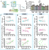
Age-dependent increase in SMOX expression correlates with oxidative stress and behavioral vulnerability following TBI. (A) Schematic overview of experimental design used to assess correlations between gene expression levels and individual behavioral or histological outcomes post-TBI. (B) Quantitative analysis of mRNA expression levels of enzymatic regulators of hydroxyl radical production in cortical tissues from young (Y), middle-aged (M), and aged (A) mice. Data are presented as mean ± SEM and analyzed by one-way ANOVA followed by Bonferroni’s post hoc test. * p < 0.05. (CE) Pearson’s correlation analyses between relative expression levels of Cybb (NOX2) (C), Smox (D), and Xdh (E) with individual TBI outcomes, including motor function (NSS), cognitive performance (ORM), and oxidative stress marker levels (4-HNE-positive cells). n = 10 mice. Small circles represent individual biological replicates. Different colors correspond to distinct experimental groups or treatment conditions.
Figure 3
Figure 3
SMOX expression is elevated in aged mouse and human brains. (A,B) Representative immunofluorescence images, showing SMOX expression (magenta) in cortical region (A) and hippocampal CA1 region (B) of young and aged mice with or without CCI surgery. Nuclei were counterstained with DAPI (white). Scale bar = 20 μm. (C,D) Quantification of SMOX fluorescence intensity in cortex (C) and hippocampal CA1 region (D) across experimental groups. (E) Quantitative analysis of SMOX mRNA levels in human frontal cortex samples across different age groups using publicly available GEO dataset GDS5204. (F,G) Representative immunofluorescence images (F) and quantification (G) of SMOX (red) and NeuN (cyan) co-staining in postmortem human frontal cortical tissue from young and aged individuals. Nuclei were counterstained with DAPI (blue). Scale bar = 20 μm. Small circles in C–E and G represent individual data points from biological replicates. All data are presented as mean ± SEM and were analyzed using one-way ANOVA followed by Bonferroni’s post hoc test. * p < 0.05.
Figure 4
Figure 4
Pharmacological inhibition of SMOX reduces oxidative stress in aged brain following TBI. (A) Schematic overview of experimental design used to evaluate effects of SMOX inhibition on oxidative stress in aged mice after CCI. (B) Representative immunofluorescence images of oxidative stress markers 4-HNE (magenta) and 8-oxo-2′-deoxyguanosine (8-oxo-dG, yellow) in hippocampal CA1 region. Nuclei are counterstained with DAPI (blue). Scale bar = 20 μm. (C,D) Quantification of 4-HNE-positive neurons (C) and 8-oxo-dG-positive cells (D) across experimental groups. Quantification based on 12 images per group (n = 4 mice/group). (E,F) Biochemical quantification of hydroxyl free radical production (E) and malondialdehyde (MDA) concentrations (F) in cortical tissues from aged mice following CCI with or without SMOX inhibition. Small circles in C–F represent individual data points from biological replicates. All data are presented as mean ± SEM and were analyzed using one-way ANOVA followed by Bonferroni’s post hoc test. * p < 0.05; ns, not significant.
Figure 5
Figure 5
Pharmacological inhibition of SMOX attenuates TBI-induced behavioral deficits in aged mice. (A) Schematic overview of experimental design used to assess effects of SMOX inhibition on functional outcomes in aged mice following CCI. (B) Quantification of motor function impairments, measured by Neurological Severity Score (NSS) and grip strength, in vehicle- and JNJ-9350-treated mice. (C) Cognitive performance assessed using Y-maze spontaneous alternation test and novel object recognition memory (ORM) test. (D) Emotional behavior evaluated through open-field center zone exploration and latency to feed in novelty-suppressed feeding (NSF) test. All data are presented as mean ± SEM and analyzed using one-way ANOVA followed by Bonferroni’s post hoc test. * p < 0.05.
Figure 6
Figure 6
SMOX expression is primarily upregulated in neurons and microglia in aged brain. (A) Representative immunofluorescence images showing co-localization of SMOX (magenta) with NeuN (neuronal marker, cyan) and Iba1 (microglial marker, green) in hippocampal CA1 region. Scale bar = 20 μm. (B) Representative images showing co-staining of SMOX (magenta) with CD31 (endothelial marker, cyan) and GFAP (astrocytic marker, green) in hippocampal CA1 region. Scale bar = 20 μm. (C) High-magnification views of representative inset areas from (A,B), illustrating detailed cellular co-localization. Scale bar = 10 μm. (D) Quantification of percentage of SMOX-positive cells co-expressing cell-type-specific markers in aged mice. Data are presented as mean ± SEM.
Figure 7
Figure 7
Pharmacological inhibition of SMOX reduces pro-inflammatory microglial activation in aged mice. (A) Representative immunofluorescence images showing co-staining of pro-inflammatory marker CD16/32 (green) and microglial marker Iba1 (magenta) in hippocampal CA1 region. Nuclei are counterstained with DAPI (blue). Scale bar = 10 μm. (B) Quantification of relative fluorescence intensity of CD16/32 across experimental groups. Quantification based on 12 images per group (n = 4 mice/group). (CF) Quantitative analysis of mRNA expression levels of canonical pro-inflammatory mediators (Il6, Il1b, Tnf, and Cxcl1) in cortical tissue following CCI, with or without JNJ-9350 treatment. All data are presented as mean ± SEM and were analyzed using one-way ANOVA followed by Bonferroni’s post hoc test. * p < 0.05; ns, not significant.
Figure 8
Figure 8
Pharmacological inhibition of SMOX reduces neuronal degeneration and apoptotic signaling in the aged brain following TBI. (A) Representative immunofluorescence images showing co-staining of degenerating neurons with Fluoro-Jade C (FJC, green) and neuronal marker NeuN (red) in hippocampal CA1 region. Nuclei are counterstained with DAPI (blue). Scale bar = 20 μm. (B) Quantification of FJC-positive neurons across experimental groups. Quantification based on 12 images per group (n = 4 mice/group). (CF) Quantitative real-time PCR analysis of mRNA expression levels of pro-apoptotic genes (Bax and Bak1) and anti-apoptotic genes (Bcl2 and Bcl2l1) in cortical tissue following CCI, with or without JNJ-9350 treatment. Small circles in B and C–F represent individual data points from biological replicates. All data are presented as mean ± SEM and analyzed using one-way ANOVA followed by Bonferroni’s post hoc test. * p < 0.05; ns, not significant.
Figure 9
Figure 9
SMOX inhibition restores GLT-1 expression and astrocytic transporter localization in aged brain following TBI. (AC) Quantitative PCR analysis of glutamate transporter gene expression in cortex of aged mice across experimental groups. CCI significantly decreased Slc1a2 (GLT-1) mRNA expression, which was reversed by SMOX inhibition (B), while Slc1a3 (EAAT1) (A) and SLC7A11 (xCT) (C) levels remained unchanged. (D) Representative immunofluorescence images showing co-localization of GFAP (green) and GLT-1 (magenta) in cortical astrocytes. Nuclei are counterstained with DAPI (blue). Scale bar = 10 μm. Dashed lines in merged images indicate regions used for line-plot quantification. (E) Line-plot analysis of GLT-1 fluorescence intensity across astrocytic regions from (D). Yellow-shaded areas represent membrane regions used to calculate membrane-to-cytosol ratios. (F) Quantification of GLT-1 signal in GFAP-positive astrocytes across experimental groups. (G) Quantification of membrane-to-cytosol GLT-1 ratio in astrocytes. CCI significantly reduced astrocytic membrane-localized GLT-1 in aged mice, effect reversed by JNJ-9350 treatment. Data are presented as mean ± SEM and were analyzed using one-way ANOVA followed by Bonferroni’s post hoc test. * p < 0.05.
Figure 10
Figure 10
SMOX inhibition with JNJ-9350 enhances long-term recovery from TBI in aged mice. (A) Schematic overview of experimental design used to assess long-term effects of SMOX inhibition on functional and histological outcomes in aged mice following CCI. (B,C) Quantification (B) and representative images (C) of Nissl-positive surviving neurons in the hippocampal CA1 region 30 days after CCI surgery, with or without JNJ-9350 treatment. (D) Quantification of motor function, assessed by Neurological Severity Score (NSS) and grip strength testing, in vehicle- and JNJ-9350-treated mice. (E) Cognitive performance measured by Y-maze spontaneous alternation test and novel object recognition memory (ORM) test. (F) Emotional behavior assessed through open-field center zone exploration and latency to feed in novelty-suppressed feeding (NSF) test. All data are presented as mean ± SEM and were analyzed using one-way ANOVA followed by Bonferroni’s post hoc test. * p < 0.05.
Figure 11
Figure 11
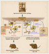
Cartoon illustration summarizing key findings of current study. In aged brain, SMOX is pathologically upregulated in neurons and microglia, leading to increased oxidative stress, neuro-inflammation, and neuronal degeneration following traumatic brain injury. In addition, SMOX dysregulation indirectly impairs astrocytic glutamate uptake by disrupting GLT-1 membrane localization, contributing to excitotoxicity. Pharmacological inhibition of SMOX using JNJ-9350 attenuates these pathological processes, resulting in reduced microglial activation, preserved neuronal integrity, and improved motor, cognitive, and emotional outcomes. These findings position SMOX as therapeutic target for mitigating age-related vulnerability to TBI.

References

    1. Ma Z., He Z., Li Z., Gong R., Hui J., Weng W., Wu X., Yang C., Jiang J., Xie L., et al. Traumatic brain injury in elderly population: A global systematic review and meta-analysis of in-hospital mortality and risk factors among 2.22 million individuals. Ageing Res. Rev. 2024;99:102376. doi: 10.1016/j.arr.2024.102376. - DOI - PubMed
    1. Gavrila Laic R.A., Bogaert L., Vander Sloten J., Depreitere B. Functional outcome, dependency and well-being after traumatic brain injury in the elderly population: A systematic review and meta-analysis. Brain Spine. 2021;1:100849. doi: 10.1016/j.bas.2021.100849. - DOI - PMC - PubMed
    1. Prasad G.L., Anmol N., Menon G.R. Outcome of Traumatic Brain Injury in the Elderly Population: A Tertiary Center Experience in a Developing Country. World Neurosurg. 2018;111:e228–e234. doi: 10.1016/j.wneu.2017.12.034. - DOI - PubMed
    1. Flanagan S.R., Hibbard M.R., Riordan B., Gordon W.A. Traumatic brain injury in the elderly: Diagnostic and treatment challenges. Clin. Geriatr. Med. 2006;22:449–468. doi: 10.1016/j.cger.2005.12.011. - DOI - PubMed
    1. Karaboue M.A.A., Ministeri F., Sessa F., Nannola C., Chisari M.G., Cocimano G., Di Mauro L., Salerno M., Esposito M. Traumatic Brain Injury as a Public Health Issue: Epidemiology, Prognostic Factors and Useful Data from Forensic Practice. Healthcare. 2024;12:2266. doi: 10.3390/healthcare12222266. - DOI - PMC - PubMed

LinkOut - more resources