Plant-Derived Nanovesicles from Soaked Rice Water: A Novel and Sustainable Platform for the Delivery of Natural Anti-Oxidant γ-Oryzanol
- PMID: 40563350
- PMCID: PMC12189669
- DOI: 10.3390/antiox14060717
Plant-Derived Nanovesicles from Soaked Rice Water: A Novel and Sustainable Platform for the Delivery of Natural Anti-Oxidant γ-Oryzanol
Abstract
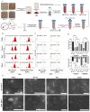
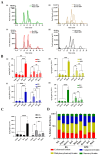
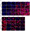
Gamma oryzanol (GO) is a natural anti-oxidant found in rice bran with potential health benefits. Conventional isolation of GO from rice bran requires the use of non-eco-friendly solvents such as acetone, ethyl acetate and hexane due to its low aqueous solubility. Further, nanoencapsulation of GO is required for the enhancement of stability and bioavailability. Plant-derived nanovesicles (PDNVs) are natural/intrinsic exosome-mimetic vesicles isolated from edible plants using green methods. Washed/soaked rice water (SRW) is often discarded as waste prior to cooking rice. However, traditional knowledge indicates its health-promoting anti-oxidant benefit, probably contributed by the presence of GO. Herein, for the first time, we isolated PDNVs from SRW by the cost-effective Polyethylene glycol 6000(PEG) precipitation method and demonstrated the presence of GO in PDNVs. In our initial screen, PDNVs were isolated from both rice grains (RGs) as well as the SRW of four different rice varieties, in which we identified the copious presence of GO in black RGs and brown SRW PDNVs. Both RG and SRW PDNVs were non-toxic to keratinocytes. SRW PDNVs displayed distinct cellular uptake mechanisms compared to RG PDNVs in human keratinocytes. Compared to native GO, brown SRW PDNVs containing GO displayed superior anti-oxidant activity in HaCaT keratinocytes, likely due to its enhanced cellular uptake. Overall, we describe here a waste-to-wealth green approach using an economical PEG method for the extraction of GO in bioavailable form. Given that oxidative stress is a driving factor for inflammation and related diseases, SRW PDNVs provide an affordable natural formulation for the treatment of diseases with underlying oxidative stress and inflammation.
Keywords: anti-oxidant activity; gamma oryzanol; plant-derived exosome-like nanovesicles; rice water.
Conflict of interest statement
The authors declare no competing interests.
Figures


Similar articles
-
Signs and symptoms to determine if a patient presenting in primary care or hospital outpatient settings has COVID-19.Cochrane Database Syst Rev. 2022 May 20;5(5):CD013665. doi: 10.1002/14651858.CD013665.pub3. Cochrane Database Syst Rev. 2022. PMID: 35593186 Free PMC article.
-
Maternal and neonatal outcomes of elective induction of labor.Evid Rep Technol Assess (Full Rep). 2009 Mar;(176):1-257. Evid Rep Technol Assess (Full Rep). 2009. PMID: 19408970 Free PMC article.
-
Carbamazepine versus phenytoin monotherapy for epilepsy: an individual participant data review.Cochrane Database Syst Rev. 2017 Feb 27;2(2):CD001911. doi: 10.1002/14651858.CD001911.pub3. Cochrane Database Syst Rev. 2017. Update in: Cochrane Database Syst Rev. 2019 Jul 18;7:CD001911. doi: 10.1002/14651858.CD001911.pub4. PMID: 28240353 Free PMC article. Updated.
-
Systemic pharmacological treatments for chronic plaque psoriasis: a network meta-analysis.Cochrane Database Syst Rev. 2021 Apr 19;4(4):CD011535. doi: 10.1002/14651858.CD011535.pub4. Cochrane Database Syst Rev. 2021. Update in: Cochrane Database Syst Rev. 2022 May 23;5:CD011535. doi: 10.1002/14651858.CD011535.pub5. PMID: 33871055 Free PMC article. Updated.
-
Systemic pharmacological treatments for chronic plaque psoriasis: a network meta-analysis.Cochrane Database Syst Rev. 2017 Dec 22;12(12):CD011535. doi: 10.1002/14651858.CD011535.pub2. Cochrane Database Syst Rev. 2017. Update in: Cochrane Database Syst Rev. 2020 Jan 9;1:CD011535. doi: 10.1002/14651858.CD011535.pub3. PMID: 29271481 Free PMC article. Updated.
References
-
- Failla M.L., Chitchumroonchokchai C., Ariyapitipun T., Adisakwattana S., Maekynen K. Effect of gamma-oryzanol on the bioaccessibility and synthesis of cholesterol. Eur. Rev. Med. Pharmacol. Sci. 2012;16:49–56. - PubMed
-
- Pan Y., Cai L., He S., Zhang Z. Pharmacokinetics study of ferulic acid in rats after oral administration of γ-oryzanol under combined use of Tween 80 by LC/MS/MS. Eur. Rev. Med. Pharmacol. Sci. 2014;18:143–150. - PubMed
Grants and funding
LinkOut - more resources
Full Text Sources

