Review
. 2025 Jun 13;14(6):726.
doi: 10.3390/antiox14060726.

Oxidized Low-Density Lipoprotein as a Potential Target for Enhancing Immune Checkpoint Inhibitor Therapy in Microsatellite-Stable Colorectal Cancer

Affiliations
Review

Oxidized Low-Density Lipoprotein as a Potential Target for Enhancing Immune Checkpoint Inhibitor Therapy in Microsatellite-Stable Colorectal Cancer

Xiaochun Zhang et al. Antioxidants (Basel). .

Abstract

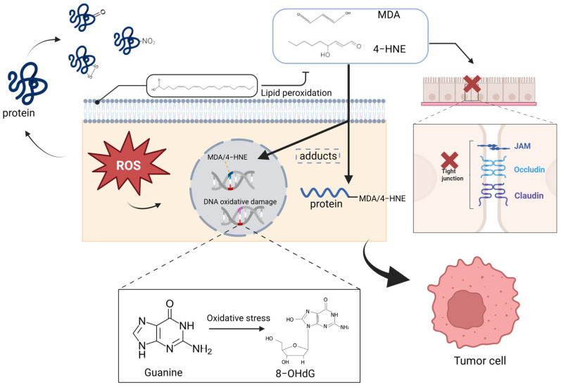
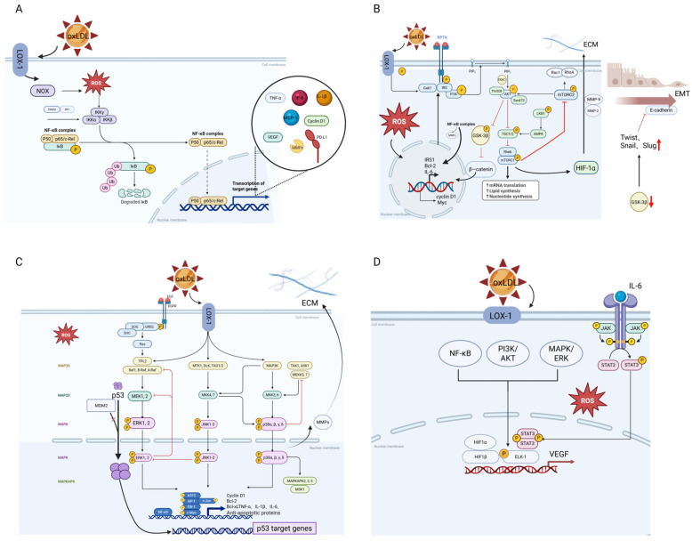
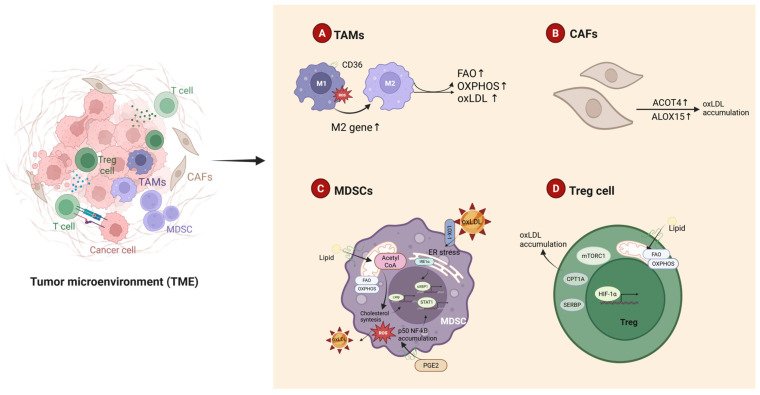
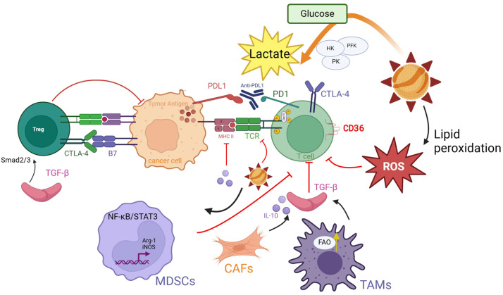
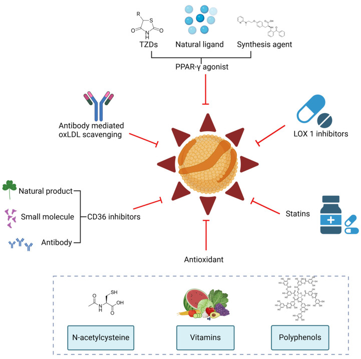
Oxidized low-density lipoprotein (oxLDL) exhibits differential expression in microsatellite-stable (MSS) and microsatellite instability-high (MSI) colorectal cancer (CRC), highlighting its potential therapeutic role in immune checkpoint inhibitor (ICI) resistance in MSS CRC. Elevated oxLDL levels in MSS CRC contribute to tumor progression and diminish ICI efficacy by modulating metabolic reprogramming and immunosuppressive mechanisms within the tumor microenvironment (TME) by activating receptors such as LOX-1 and CD36. oxLDL triggers signaling pathways, including NF-κB, PI3K/Akt, and MAPK, leading to the expansion of immunosuppressive cells like regulatory T cells (Tregs), myeloid-derived suppressor cells (MDSCs), and M2 macrophages, while concurrently suppressing effector T cell functions. Additionally, oxLDL enhances oxidative stress and promotes fatty acid oxidation (FAO) and glycolytic metabolism, resulting in nutrient competition within the TME and establishing an immunosuppressive milieu, ultimately culminating in ICI resistance. This review systematically examines the disparities in oxLDL expression between MSS and MSI CRC and elucidates the molecular mechanisms through which oxLDL mediates ICI resistance. Furthermore, it explores potential therapeutic strategies targeting oxLDL, offering novel avenues to overcome immunotherapy resistance in MSS CRC.

Keywords: immune checkpoint inhibitors; immunosuppression; metabolic reprogramming; microsatellite-stable colorectal cancer; oxidative stress; oxidized low-density lipoprotein; tumor microenvironment.


Conflict of interest statement

The authors declare no conflicts of interest.

Figures

Figure 1
Figure 1
Mechanisms of ROS generation: endogenous and exogenous sources. Endogenous sources include mitochondrial electron transport chain activity, NADPH oxidase (NOX) enzymes, peroxisomal metabolism, cytochrome P450 enzyme activity, xanthine oxidoreductase (XOR), nitric oxide synthase (NOS, including nNOS, iNOS, and eNOS), and cyclooxygenase/lipoxygenase (COX/LOX). Exogenous sources encompass ultraviolet (UV) radiation, ionizing radiation, environmental toxins, and certain chemotherapeutic agents. Created in BioRender.com.
Figure 2
Figure 2
OS causes oxidative damage in DNA, lipids, and proteins, promoting the initiation and metastasis of CRC. ROS can cause DNA base oxidation and strand breaks, leading to genomic instability and mutations that promote tumorigenesis. They also react with polyunsaturated fatty acids in cell membranes, producing lipid peroxides like malondialdehyde (MDA) and 4-hydroxynonenal (4-HNE). This process disrupts membrane fluidity and integrity, increases permeability, and indirectly impairs tight junctions, further promoting ROS generation. Additionally, ROS oxidize amino acid residues in proteins, resulting in carbonylation and nitration, impairing functions of DNA repair enzymes like OGG1 and tumor suppressors such as p53, thereby accelerating cancer progression. Created in BioRender.com.
Figure 3
Figure 3
Expression pathways of oxLDL in CRC. (A) NF-κB Inflammatory Signaling: oxLDL binds to LOX-1, activating IKK and releasing NF-κB, which upregulates pro-inflammatory factors (e.g., TNF-α, IL-6) and immune checkpoint molecules (e.g., PD-L1), driving inflammation and immune evasion. (B) PI3K/Akt Signaling: oxLDL activates PI3K/Akt, enhancing cell survival, proliferation, and metabolic reprogramming by inhibiting apoptosis and upregulating anti-apoptotic genes (e.g., Bcl-2). Inactivation of GSK-3β enhances the expression of EMT-related factors (e.g., Twist, Snail) and suppresses the expression of the cell adhesion molecule E-cadherin, promoting cancer cell invasion and migration. (C) MAPK Signaling: oxLDL triggers ERK1/2, p38, and JNK pathways, promoting cell proliferation, invasion, and metastasis via transcription factors (e.g., AP-1). (D) VEGF signaling: oxLDL upregulates VEGF through NF-κB, PI3K/Akt, and MAPK, stimulating angiogenesis and immune evasion by enhancing endothelial cell proliferation and vascular permeability. Created in BioRender.com.
Figure 4
Figure 4
Differential accumulation of oxLDL in the immune microenvironment of MSS CRC. (A) TAMs: oxLDL is internalized by TAMs via LOX-1/CD36 receptors, enhancing FAO and OXPHOS, driving M2 polarization, and facilitating oxLDL uptake and intracellular accumulation. (B) CAFs: CAFs upregulate CD36 and LOX-1 expression through the secretion of IL-6 and TGF-β, promoting oxLDL uptake. ALOX15 and ACOT4 accelerate lipid peroxidation, leading to abnormal oxLDL accumulation. (C) MDSCs: MDSCs enhance FAO and OXPHOS, uptake oxLDL, and generate excessive ROS, inducing endoplasmic reticulum stress and suppressing T cell function. (D) Tregs: Tregs undergo lipid uptake and metabolic reprogramming mediated by CD36 and PPARγ, while the PD-1/CPT1A axis enhances FAO, further suppressing effector T cell activity. Collectively, these mechanisms contribute to the abnormal accumulation of oxLDL in the TME of MSS-type CRC, establishing a positive feedback loop that reinforces immune suppression and tumor progression. Created in BioRender.com.
Figure 5
Figure 5
OxLDL-mediated resistance mechanisms to ICIs. (1) MDSCs: oxLDL activates the NF-κB/STAT3 axis in MDSCs, inducing overexpression of Arg-1 and iNOS, which deplete arginine and generate reactive nitrogen species, establishing a metabolic immunosuppressive barrier. (2) CAFs: oxLDL enhances CAF-mediated secretion of TGF-β and IL-10, fostering stromal fibrosis and physical barriers that impede T cell infiltration. CAFs further amplify immunosuppression by recruiting MDSCs and Tregs via chemokine signaling. (3) TAMs: oxLDL drives M2 polarization of TAMs through CD36/PPARγ signaling, enhancing their glycolytic flux and lactate production. Lactate activates the MCT1/NF-κB/COX-2 axis to upregulate PD-L1 on neutrophils and PD-1 on Tregs, while TME acidification directly suppresses cytotoxic T cell activity. (4) Metabolic Reprogramming Synergy: oxLDL upregulates glycolysis enzymes (e.g., HK, PKM) in tumor cells and TAMs, depleting glucose and inducing T cell energy exhaustion. Concurrently, oxLDL activates FAO via CD36/CPT1A, fueling immunosuppressive functions of TAMs and MDSCs. Created in BioRender.com.
Figure 6
Figure 6
Potential therapeutic strategies targeting oxLDL-mediated immunosuppression. (1) Antibody-mediated oxLDL scavenging: The E06 monoclonal antibody specifically binds to oxidized phospholipid epitopes on oxLDL, inhibiting the oxLDL-TLR4/CD36 signaling axis. (2) LOX-1 and (3) CD36 Inhibitors: These agents block oxLDL internalization and downstream signaling. (4) Antioxidants: N-acetylcysteine and polyphenolic compounds scavenge ROS, reduce oxLDL generation, and restore immune cell function. (5) Metabolic Modulation: Statins inhibit HMG-CoA reductase, lowering cholesterol synthesis and oxLDL levels. (6) PPAR-γ Agonists: These agents upregulate ABCA1/ABCG1 to promote cholesterol reverse transport while suppressing the NF-κB pathway, alleviating inflammation and immune suppression. These multi-target strategies hold promise for reversing oxLDL-mediated immune suppression and enhancing ICI efficacy. Created in BioRender.com.

Similar articles

References

    1. Perillo B., Di Donato M., Pezone A., Di Zazzo E., Giovannelli P., Galasso G., Castoria G., Migliaccio A. ROS in cancer therapy: The bright side of the moon. Exp. Mol. Med. 2020;52:192–203. doi: 10.1038/s12276-020-0384-2. - DOI - PMC - PubMed
    1. Snezhkina A.V., Kudryavtseva A.V., Kardymon O.L., Savvateeva M.V., Melnikova N.V., Krasnov G.S., Dmitriev A.A. ROS Generation and Antioxidant Defense Systems in Normal and Malignant Cells. Oxid. Med. Cell Longev. 2019;2019:6175804. doi: 10.1155/2019/6175804. - DOI - PMC - PubMed
    1. Foksinski M., Rozalski R., Guz J., Ruszkowska B., Sztukowska P., Piwowarski M., Klungland A., Olinski R. Urinary excretion of DNA repair products correlates with metabolic rates as well as with maximum life spans of different mammalian species. Free Radic. Biol. Med. 2004;37:1449–1454. doi: 10.1016/j.freeradbiomed.2004.07.014. - DOI - PubMed
    1. Wei S.C., Duffy C.R., Allison J.P. Fundamental Mechanisms of Immune Checkpoint Blockade Therapy. Cancer Discov. 2018;8:1069–1086. doi: 10.1158/2159-8290.CD-18-0367. - DOI - PubMed
    1. Brand M.D. The sites and topology of mitochondrial superoxide production. Exp. Gerontol. 2010;45:466–472. doi: 10.1016/j.exger.2010.01.003. - DOI - PMC - PubMed

LinkOut - more resources