. 2025 May 23;15(6):755.
doi: 10.3390/biom15060755.

Separation-of-Function Alleles of smc-5 Reveal Domain-Specific Defects and a Conserved Residue Critical for Genome Maintenance

Affiliations

Separation-of-Function Alleles of smc-5 Reveal Domain-Specific Defects and a Conserved Residue Critical for Genome Maintenance

Haiyan Yuan et al. Biomolecules. .

Abstract

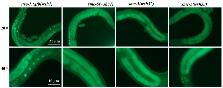
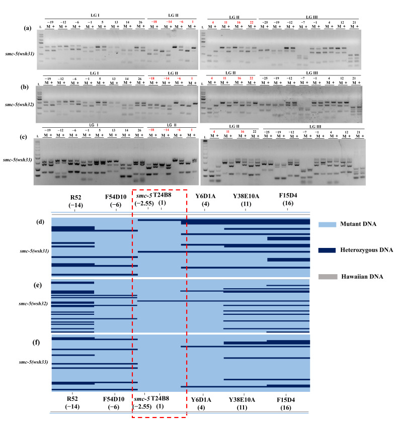
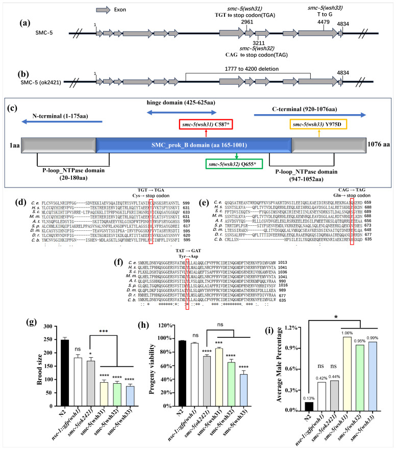
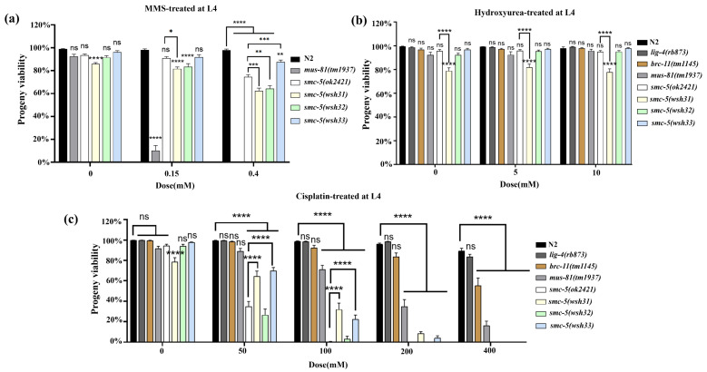
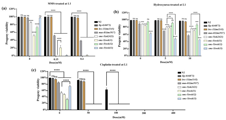
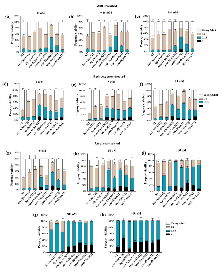
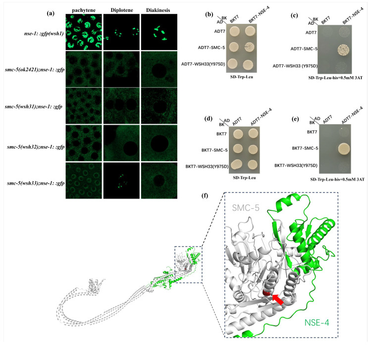
The SMC-5/6 complex safeguards genome stability through the coordinated action of its core SMC proteins and associated NSE subunits. NSE-1 is a key component of the complex and is essential for DNA repair, yet it remains poorly characterized in Caenorhabditis elegans. To further elucidate the functional mechanisms of NSE-1, we performed an EMS-based forward genetic screen in an nse-1::gfp(wsh1) reporter strain to identify mutants with defective NSE-1 expression or nuclear localization. We isolated three mutants; smc-5(wsh31), smc-5(wsh32), and smc-5(wsh33), that display impaired NSE-1::GFP nuclear localization. SNP mapping and whole-genome sequencing revealed three novel smc-5 alleles: two truncations, alleles smc-5(wsh31) (C587*) and smc-5(wsh32) (Q655*), and one missense variant, smc-5(wsh33) (Y975D), each altering a highly conserved residue in the SMC domain. All three mutants exhibited significantly reduced brood size, progeny viability, and slightly elevated male percentages. Phenotypic characterization revealed that the truncations completely abrogate NSE-1::GFP nuclear localization, whereas the missense allele causes stage-dependent, partial mislocalization. Functional assays further demonstrated allele-specific and developmental stage-dependent hypersensitivities to DNA-damaging agents (MMS, HU, and cisplatin). These separation-of-function smc-5 alleles underscore the importance of domains and conserved residues in complex integrity and genome maintenance, and provide powerful genetic tools to dissect SMC-5/6 functions in vivo.

Keywords: Caenorhabditis elegans; NSE-1 localization; SMC-5/6 complex; genome stability; smc-5.


Conflict of interest statement

The authors declare no conflicts of interest.

Figures

Figure 1
Figure 1
Mutations smc-5(wsh31), smc-5(wsh32), and smc-5(wsh33) cause aberrant NSE-1::GFP localization in the C. elegans germline. Fluorescence microscopy images comparing germline nuclei from the parental nse-1::gfp(wsh1) control strain and the indicated mutants (smc-5(wsh31), smc-5(wsh32), and smc-5(wsh33)). Representative images acquired with 20× (upper row) and 40× (lower row) objectives are shown for each genotype. In the parental control strain, NSE-1::GFP exhibits clear localization to chromosomes within the nuclei. In contrast, NSE-1::GFP signal is largely excluded from the nucleus in the smc-5(wsh31) and smc-5(wsh32) mutants. The smc-5(wsh33) mutant displays partial mislocalization, characterized by reduced chromosomal association compared to the control. Images were captured using a Leica DM6B fluorescence microscope. “wsh” is the allele designation assigned by the Caenorhabditis Genetics Center (CGC) to our lab.
Figure 2
Figure 2
Chromosome and interval mapping of mutations in smc-5(wsh31), smc-5(wsh32), and smc-5(wsh33). (ac) Chromosome mapping results for smc-5(wsh31) (a), smc-5(wsh32) (b), and smc-5(wsh33) (c). Agarose gels display DraI-digested PCR products for 48 SNPs across chromosomes I, II, III, IV, V, and X, with genetic positions shown in red (-18, -14, -4, 1, 4, 11, 16, 22) (Figures S1–S3). Lanes are labeled “M” (mutant) and “+” (wild-type). On chromosome II, mutant lanes exhibit enrichment of Bristol-specific bands, indicating linkage, whereas other chromosomes show identical patterns between mutant and wild-type lanes. (df) Interval mapping results for smc-5(wsh31) (d), smc-5(wsh32) (e), and smc-5(wsh33) (f) on chromosome II. Each row represents an individual recombinant, and each column corresponds to a chromosome II SNP. Blue indicates homozygous Bristol (N2), purple indicates heterozygous (N2/CB4856), and grey indicates homozygous Hawaiian (CB4856). The red dashed rectangle delineates the region of minimal recombination, indicative of tight linkage to the mutations.
Figure 3
Figure 3
Identification and characterization of smc-5 mutations in smc-5(wsh31), smc-5(wsh32), and smc-5(wsh33). (a) Schematic of the smc-5 gene structure on chromosome II, showing the positions of the mutations in smc-5(wsh31) (C587*), smc-5(wsh32) (Q655*), and smc-5(wsh33) (Y975D). Exons are depicted as boxes, with the SMC domain highlighted in grey. (b) Comparison with the smc-5(ok2421) deletion allele, which removes a large portion of the SMC domain. (c) Mapping of the mutations onto the SMC-5 protein, showing their positions within the SMC domain (Figure S4b). (df) Amino acid sequence alignments of SMC-5 regions across C. elegans, H. sapiens, S. cerevisiae (yeast), M. musculus, A. thaliana, S. pombe, D. melanogaster, D. rerio, and C. briggsae. Red vertical rectangular boxes highlight the position of the amino acid residue affected. (g) Brood size of N2, nse-1::gfp(wsh1), smc-5(ok2421), smc-5(wsh31), smc-5(wsh32), and smc-5(wsh33) mutants, showing significant reductions in all mutants compared to N2. (h) Progeny viability, measured as the percentage of viable offspring, with all mutants showing reduced viability compared to N2. (i) Male frequency, indicating increased X-chromosome nondisjunction in all the new mutants compared to N2, nse-1::gfp(wsh1), and smc-5(ok2421). Data are presented as mean ± SEM; statistical significance was determined by Student’s t-test (* p < 0.05, *** p < 0.001, **** p < 0.0001, ns: not significant). Number of animals n; N2 = 28, nse-1::gfp(wsh1) = 25, smc-5(ok2421) = 28, smc-5(wsh31) = 25, smc-5(wsh32) = 22, smc-5(wsh33) = 24 (Table S1). “wsh” is the allele designation assigned by the CGC to our lab.
Figure 4
Figure 4
Differential sensitivities of smc-5 alleles to genotoxic agents. (a) Viability assays of L4-stage C. elegans exposed to varying concentrations of methyl methanesulfonate (MMS). (b) Viability assays of L4-stage worms subjected to replication stress induced by hydroxyurea (HU). (c) Viability assays of L4-stage worms exposed to escalating concentrations of cisplatin, which induces inter- and intra-strand DNA crosslinks. Bars represent the mean ± SEM for each genotype, based on three independent biological replicates. Statistical significance was evaluated using one-way ANOVA with multiple-comparison corrections. Asterisks indicate levels of significance relative to N2 or the appropriate control group, as detailed in the text (* p < 0.05, ** p < 0.01, *** p < 0.001, **** p < 0.0001, ns: not significant). Sample sizes (n) are available in Tables S2–S4 for (ac), respectively. No significant difference was observed between N2 and nse-1::gfp(wsh1), indicating that the GFP tag did not affect the assay outcome (Figure S5).
Figure 5
Figure 5
DNA damage sensitivity of L1-stage wild-type and DNA repair mutant C. elegans strains, measured by progeny viability. Percent progeny viability following exposure to varying doses of (a) methyl methanesulfonate (MMS; alkylating agent), (b) hydroxyurea (HU; replication stress), and (c) cisplatin (inter- and intra-strand crosslinks). Asterisks indicate levels of significance relative to N2 or the appropriate control group, as detailed in the text (** p < 0.01, *** p < 0.001, **** p < 0.0001, ns: not significant). Sample sizes (n) are available in Tables S5–S7 for (ac), respectively. No significant difference was observed between N2 and nse-1::gfp(wsh1), indicating that the GFP tag did not affect the assay outcome (Figure S6).
Figure 6
Figure 6
Developmental outcomes 48 h after L1-stage exposure to MMS, hydroxyurea, or cisplatin. Bars represent the percentages of animals reaching the indicated developmental stages at each treatment dose. Panels illustrate outcomes following exposure to (ac) MMS (0, 0.15, 0.4 mM), (df) hydroxyurea (0, 5, 10 mM), and (gk) cisplatin (0, 50, 100, 200, 400 µM). Sample sizes (n) are available in Tables S8–S10 for (ak), respectively. No unusual difference was observed between N2 and nse-1::gfp(wsh1), indicating that the GFP tag did not affect the assay outcome (Figure S7).
Figure 7
Figure 7
NSE-1::GFP localization depends on the SMC-5–NSE-4 interaction. (a) High-resolution fluorescence micrographs of C. elegans germline nuclei (pachytene, diplotene, and diakinesis stages) showing NSE-1::GFP localization in control and smc-5 mutant backgrounds (scale bars, 10 μm). (b) Yeast two-hybrid (Y2H) control plate (SD –Trp –Leu) showing growth of all transformants, confirming they carry both SMC-5 and NSE-4 plasmids. (c) Y2H interaction test (SD –Trp –Leu –His + 3AT) with wild-type or Y975D mutant SMC-5 fused to the Gal4 activation domain (AD) and NSE-4 fused to the Gal4 DNA-binding domain (BD). (d) Y2H control plate for the reciprocal configuration (SD –Trp –Leu), where SMC-5 is in the BD vector and NSE-4 in the AD vector. (e) Selective interaction plate (SD –Trp –Leu –His + 3AT) for the reciprocal configuration. (f) The 3D model of the SMC-5/NSE-4 interface illustrating the position of the Y975D substitution.

Similar articles

References

    1. Wu N., Yu H. The Smc Complexes in DNA Damage Response. Cell Biosci. 2012;2:5. doi: 10.1186/2045-3701-2-5. - DOI - PMC - PubMed
    1. Peng X.P., Zhao X. The Multi-Functional Smc5/6 Complex in Genome Protection and Disease. Nat. Struct. Mol. Biol. 2023;30:724–734. doi: 10.1038/s41594-023-01015-6. - DOI - PMC - PubMed
    1. Serrano D., Cordero G., Kawamura R., Sverzhinsky A., Sarker M., Roy S., Malo C., Pascal J.M., Marko J.F., D’Amours D. The Smc5/6 Core Complex Is a Structure-Specific DNA Binding and Compacting Machine. Mol. Cell. 2020;80:1025–1038. doi: 10.1016/j.molcel.2020.11.011. - DOI - PMC - PubMed
    1. Pradhan B., Kanno T., Umeda Igarashi M., Loke M.S., Baaske M.D., Wong J.S.K., Jeppsson K., Björkegren C., Kim E. The Smc5/6 Complex Is a DNA Loop-Extruding Motor. Nature. 2023;616:843–848. doi: 10.1038/s41586-023-05963-3. - DOI - PMC - PubMed
    1. Yu Y., Li S., Ser Z., Kuang H., Than T., Guan D., Zhao X., Patel D.J. Cryo-EM Structure of DNA-Bound Smc5/6 Reveals DNA Clamping Enabled by Multi-Subunit Conformational Changes. Proc. Natl. Acad. Sci. USA. 2022;119:e2202799119. doi: 10.1073/pnas.2202799119. - DOI - PMC - PubMed

MeSH terms

Substances

LinkOut - more resources