. 2025 May 27;15(6):768.
doi: 10.3390/biom15060768.

Targeting Glioblastoma Stem Cells: A40s Aptamer-NIR-Dye Conjugate for Glioblastoma Visualization and Treatment

Affiliations

Targeting Glioblastoma Stem Cells: A40s Aptamer-NIR-Dye Conjugate for Glioblastoma Visualization and Treatment

Alessandra Affinito et al. Biomolecules. .

Abstract

Glioblastoma (GBM) is the most aggressive and challenging brain cancer, in terms of diagnosis and therapy. The highly infiltrative glioblastoma stem cells (GSCs) are difficult to visualize and surgically remove with the current diagnostic tools, which often lead to misdiagnosis and false-positive results. In this study, we focused on a groundbreaking tool for specifically visualizing and removing GSCs. We exploited the specific binding of A40s aptamer to EphA2 for the selective delivery of Near-Infrared Dyes (NIR-Dyes), like IR700DX and ICG, both in vitro and in vivo. The A40s aptamer, engineered through the NIR-Dye conjugation, did not affect aptamer binding ability; indeed, A40s-NIR-Dye conjugates bound GLI261 stem-like cells and patient-derived GSCs in vitro; moreover, they induced cell death upon photodynamic therapy treatment (PDT). Additionally, when systemically administrated, the A40s-NIR-Dye conjugates allowed GSC visualization and accumulated in tumor mass. This allows GSCs detection and treatment. Our findings demonstrate the potential use of A40s aptamer as a targeted therapeutic approach and imaging tool in vivo for GSCs, paving the way for improved, more effective, and less invasive GBM management.

Keywords: A40s; EphA2; GBM; ICG; IR700DX; NIR dye; PDT; aptamers; glioblastoma stem cells; guide surgery; photodynamic therapy.


Conflict of interest statement

Authors Alessandra Affinito, Sara Verde and Aurelia Fraticelli were employed by the company AKA Biotech. The remaining authors declare that the research was conducted in the absence of any commercial or financial relationships that could be construed as a potential conflict of interest.

Figures

Figure 1
Figure 1
A40s-NIR-Dye conjugates characterization. (A) Schematic representation of the reaction and the resulting aptamer-NIR-Dye conjugate. (B) Non-denaturing 12% acrylamide gel electrophoresis of A40s-IR700DX chimera and controls (non-related aptamer-based chimera, Scra-IR700DX, and non-labeled aptamers); simultaneous visualizing of the dye portion by white light irradiation (right panel) and RNA by ultraviolet (UV) light (left panel). Original images can be found in Supplementary Materials.
Figure 2
Figure 2
Binding ability and functional assay of A40s-IR700DX on GSCs. (A) Binding ability of A40s-IR700DX chimera on patient-derived GSCs. Representative experiment is shown, and result is expressed relative to the background binding detected with a scrambled chimera used as a negative control. (B) Caspase activity and (C) cell viability of patient-derived GSCs 48 h and 72 h after 400 nM A40s-IR700DX or Scra-IR700DX chimera treatment. Results are presented as mean ± SD of three replicates. **** p ≤ 0.0001.
Figure 3
Figure 3
GL261-luc2- stem-like cells as a GBM mouse model. (A) Western blot analysis of EphA2 expression in GL261 differentiated and stem-like cells (after 10 and 17 days). (B) A40s aptamer binding ability on differentiated and GL261-luc2 stem-like cells. Representative experiment is shown, and result is expressed ± SD relative to the background binding detected with a scrambled chimera used as negative control. (C) A40s-IR700DX chimera binding ability after 15 and 30 min of incubation with GL261 stem-like cells evaluated by fluorescence scanning (Odyssey CLx Imager). Representative experiment is shown, and result is expressed ± SD relative to the background binding detected with a scrambled chimera used as negative control. (D) Cell viability of GL261 stem-like cells (MTS assay) upon 10 J/cm2 after 1 h treatment with A40s-IR700DX chimera and NIR-dye free. Results are presented as mean ± SD of three replicates. * p ≤ 0.05; ** p ≤ 0.01; *** p ≤ 0.001. Original images can be found in Supplementary Materials.
Figure 4
Figure 4
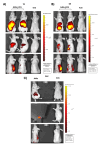
A40s-ICG chimera in vivo biodistribution on GL261-luc2 stem-like cells engrafted into BALB\C nude mice. A40s-ICG chimera and ICG free clearance evaluation after 1 h (A), 6 h (B), and 24 h (C) treatments by IVIS Spectrum imaging system.
Figure 5
Figure 5
A40s-ICG chimera ex vivo biodistribution in GL261-luc2 stem-like cells engrafted into BALB\C nude mice. A40-ICG ex vivo detection in organs by IVIS Spectrum imaging system after 6 h (A) and 24 h (B) treatments; (C) organs of control mouse treated with free ICG after 24 h of treatment.

References

    1. Sabouri M., Dogonchi A.F., Shafiei M., Tehrani D.S. Survival rate of patient with glioblastoma: A population-based study. Egypt. J. Neurosurg. 2024;39:42. doi: 10.1186/s41984-024-00294-5. - DOI
    1. Eckerdt F., Platanias L.C. Emerging Role of Glioma Stem Cells in Mechanisms of Therapy Resistance. Cancers. 2023;15:3458. doi: 10.3390/cancers15133458. - DOI - PMC - PubMed
    1. Seker-Polat F., Pinarbasi Degirmenci N., Solaroglu I., Bagci-Onder T. Tumor Cell Infiltration into the Brain in Glioblastoma: From Mechanisms to Clinical Perspectives. Cancers. 2022;14:443. doi: 10.3390/cancers14020443. - DOI - PMC - PubMed
    1. Vollmann-Zwerenz A., Leidgens V., Feliciello G., Klein C.A., Hau P. Tumor Cell Invasion in Glioblastoma. Int. J. Mol. Sci. 2020;21:1932. doi: 10.3390/ijms21061932. - DOI - PMC - PubMed
    1. Wykosky J., Gibo D.M., Stanton C., Debinski W. EphA2 as a novel molecular marker and target in glioblastoma multiforme. Mol. Cancer Res. 2005;3:541–551. doi: 10.1158/1541-7786.MCR-05-0056. - DOI - PubMed