. 2025 Jun 4;15(6):817.
doi: 10.3390/biom15060817.

Enhanced Innate Immunity Mediated by IL-36α in Atopic Dermatitis and Differences in Cytokine Profiles of Lymphocytes in the Skin and Draining Lymph Nodes

Affiliations

Enhanced Innate Immunity Mediated by IL-36α in Atopic Dermatitis and Differences in Cytokine Profiles of Lymphocytes in the Skin and Draining Lymph Nodes

Ayaka Ichikawa et al. Biomolecules. .

Abstract

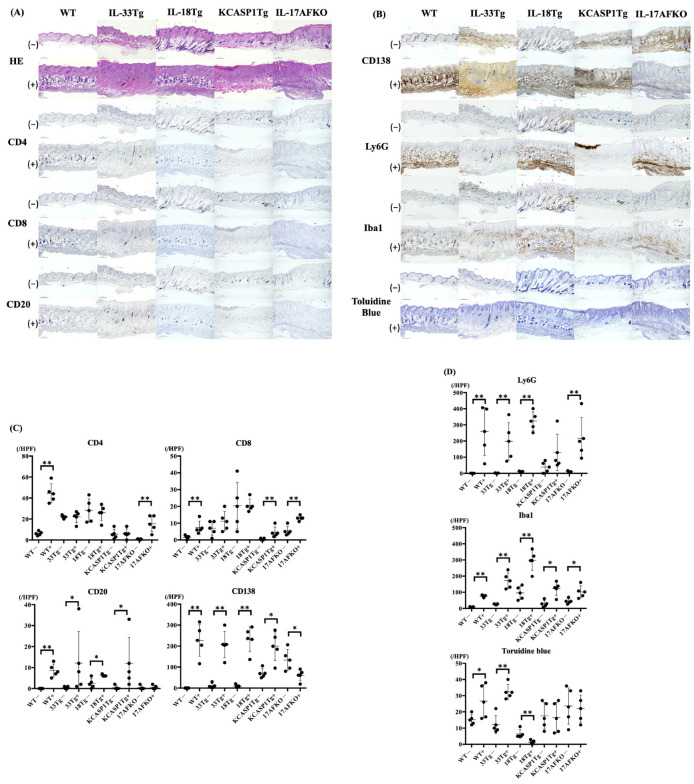
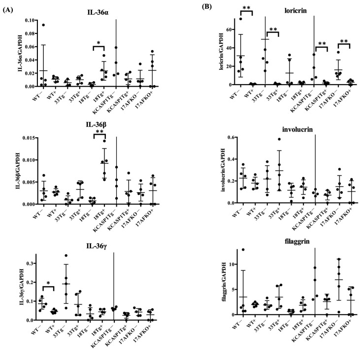
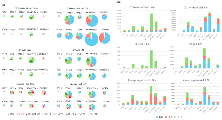
(1) Background: The IL-36 cytokines have been identified as key contributors to pustular psoriasis, and their inhibitor is already in clinical use. However, few studies have explored them in atopic dermatitis. (2) Methods: The role of IL-36α was investigated in various atopic dermatitis models using wild-type, keratin 14-specific IL-33 transgenic, IL-18 transgenic, caspase-1 transgenic, and caspase-1 transgenic mice with IL-17AF deletion, reflecting diverse aspects of human skin inflammation. IL-36α was administered subcutaneously in five doses on alternate days across the five strains to examine cellular infiltration patterns and cytokine expression levels. (3) Results: The skin phenotype was exacerbated, accompanied by worsening edema and skin thickness in all mouse groups upon IL-36α administration. An increase in infiltrating cells was observed among innate immune cells, while lymphocyte counts, including T cells and innate lymphoid cells, did not rise. Additionally, anti-inflammatory cytokines were induced simultaneously with inflammatory cytokines and downstream cytokines of IL-36α as well. Infiltrating lymphocytes in the skin displayed a distinct Type 2 cytokine-dominant profile for innate lymphoid cells and a Type 3 cytokine-dominant profile for T helper cells and γδ T cells, contrasting with the Type 1-dominant cell profile in draining lymph nodes. Type 1, Type 2, and Type 3 cytokine dominance patterns were not affected by the administration of IL-36α. (4) Conclusions: IL-36α triggers inflammatory responses in atopic dermatitis by activating innate immunity. The infiltrating lymphocytes in the skin have different cytokine production profiles between innate lymphoid cells and T cells, as well as different patterns of cytokine production in their draining lymph nodes.

Keywords: alarmin; atopic dermatitis; inflammation; interleukin-36α; psoriasis.


Conflict of interest statement

The authors declare no conflicts of interest. The funders had no role in the design of this study; in the collection, analysis, or interpretation of data; in the writing of this manuscript; or in the decision to publish these results.

Figures

Figure 1
Figure 1
(A) Clinical features of WT, IL-33Tg, IL-18Tg, KCASP1Tg, and KCASP1Tg+IL-17AFKO mice, with or without IL-36α treatment. “(–)” indicates the IL-36α-untreated group; “(+)” indicates the IL-36α-treated group. WT mice did not develop skin eruptions under baseline conditions; however, IL-36α injection induced cutaneous inflammation, characterized by mild erosions and lichenification, particularly around the eyes and nose. In IL-33Tg mice, erythema with exudation, erosions, crusts, scaling, and alopecia were observed around the periorbital and perinasal regions, as well as the ears, even without IL-36α treatment. Upon IL-36α administration, IL-33Tg mice exhibited exacerbated skin thickening, erosions, and edema. In IL-18Tg mice, a chronic AD model, severe erosive dermatitis, reepithelialization, and lichenoid inflammation were evident. IL-36α treatment further induced pronounced lichenification and crusting. Skin thickening and loss of elasticity rendered eye opening difficult in some mice. In KCASP1Tg mice without IL-36α, erosions originated on the face and spread to the ears and neck. Multiple ulcers developed on the face, trunk, and extremities. Although reepithelialization with atrophic skin occurred, erosions and ulcers frequently relapsed. Facial and eyelid hair was lost, and a fibrotic membrane covered the eyes. In IL-36α-treated KCASP1Tg mice, erosions enlarged to cover the entire face, accompanied by pronounced scarring and skin thickening. KCASP1Tg mice lacking IL-17AF (KCASP1Tg+IL-17AFKO) developed milder inflammation than KCASP1Tg mice. Upon IL-36α treatment, these mice exhibited localized erosions and crusts, predominantly around the eyes and face. Representative images of mice from each group are shown. (B) The area of facial and neck skin erosion and the ulcer was calculated using ImageJ JS. The percentage of area of skin lesions was increased in all groups when IL-36α was administered, compared to the non-treated group. The area of skin lesions was larger in the KCASP1Tg group than in the other groups (** p < 0.01).
Figure 2
Figure 2
(A) H&E and CD4, CD8, CD20 immunohistochemical stainings of skin tissue from WT, IL-33Tg, IL-18Tg, KCASP1Tg, and KCASP1Tg+IL-17AFKO mice, observed at 10× magnification (scale bar 200 μm). All strains of mice, but not the WT group, showed large infiltration of immune cells even in IL-36α non-treated groups. After IL-36α administration, all mouse models exhibited massive immune cell infiltration and epidermal thickening. (B) CD138, Ly6G, and Iba1 immunohistochemical staining and toluidine blue staining of skin tissue from the same mouse models, observed at 10× magnification (scale bar 200 μm). CD138, Ly6G, and Iba1 staining showed the presence of plasma cells, neutrophils, and monocytes or macrophages, respectively. CD138 and Ly6G positive cells were increased significantly in IL-36α-administrated groups. (C,D) Quantitative analysis of immune cell infiltration. Positive cells were counted at five randomly selected fields (400× magnification) per sample. In the IL-36α administration group, WT mice showed an increase in all types of cells, while IL-33Tg mice exhibited an increase in CD20, CD138, Ly6G, Iba1, and toluidine blue-positive cells. IL-18Tg mice demonstrated increases in CD20, CD138, Ly6G, Iba1, and toluidine blue-positive cells. KCASP1Tg mice showed an increase in CD8, CD20, CD138, and Iba1-positive cells, and KCASP1Tg+IL-17AFKO mice had an increase in CD4, CD8, CD138, Ly6G, and Iba1-positive cells. All groups were analyzed using the Mann–Whitney U test (* p < 0.05; ** p < 0.01).
Figure 3
Figure 3
(A) Expression levels of pro-inflammatory cytokines (TNFα, IFNγ, IL-4, IL-13, IL-17A, and IL-17F) were assessed in skin samples from WT, IL-33 transgenic (IL-33Tg), IL-18 transgenic (IL-18Tg), KCASP1 transgenic (KCASP1Tg), and KCASP1Tg+IL-17AF knockout (KCASP1Tg+IL-17AFKO) mice, with or without IL-36α treatment. RT-PCR analysis revealed upregulation of Type 2 cytokines, including IL-5 and IL-13, following IL-36α administration in WT mice. In contrast, Type 1 cytokines exhibited differential responses: TNFα was constitutively expressed but tended to decrease, possibly due to a shift toward Type 2 cytokine induction; IFNγ expression showed a modest increase. IL-17AF levels remained unchanged in WT mice. In IL-18Tg mice, IL-36α administration resulted in increased expression of IFNγ, IL-4, IL-5, and IL-13. No notable changes were observed in KCASP1Tg or KCASP1Tg+IL-17AFKO mice. (B) Gene expression associated with NF-κB and MAPK signaling pathways following IL-36α treatment. Within the NF-κB pathway, IL-6 expression was significantly elevated in IL-18Tg mice, while CCL2 levels increased in both WT and IL-33Tg mice. Regarding the MAPK pathway, EGR1 expression was significantly downregulated in WT mice, and c-Fos expression was reduced in WT, IL-18Tg, and KCASP1Tg+IL-17AFKO mice. MMP-9 expression was markedly upregulated in IL-18Tg mice, whereas COX-2 expression was significantly suppressed in both WT and KCASP1Tg+IL-17AFKO mice. Statistical significance was assessed using the Mann–Whitney U test (* p < 0.05; ** p < 0.01).
Figure 4
Figure 4
The expression levels of anti-inflammatory cytokines (TGFβ, IL-10, and IL-27) were measured in skin samples from the same groups of mice. TGFβ was increased in IL-33Tg after IL-36α injection. After IL-36α administration, IL-10 showed an increasing trend in whole strains, but no statistically significant increase was observed in any of the AD mouse models. IL-27 levels were increased in WT and IL-18Tg following IL-36α injection. Statistical significance was determined using the Mann–Whitney U test (* p < 0.05; ** p < 0.01).
Figure 5
Figure 5
(A) Expression levels of IL-36 isoforms (Il36a, Il36b, and Il36g) were analyzed in skin samples from WT, IL-33 transgenic (IL-33Tg), IL-18 transgenic (IL-18Tg), KCASP1 transgenic (KCASP1Tg), and KCASP1Tg+IL-17AF knockout (KCASP1Tg+IL-17AFKO) mice, with or without IL-36α treatment. Following IL-36α administration, Il36a and Il36b expression was significantly upregulated in IL-18Tg mice. In contrast, Il36g expression was significantly downregulated in WT mice and tended to decrease in the other strains. (B) Expression levels of epidermal barrier-associated genes (loricrin, involucrin, and filaggrin) were evaluated in the same set of samples. Loricrin expression was consistently reduced across all strains after IL-36α treatment. However, no statistically significant changes were observed in the expression of involucrin or filaggrin in any strain. Statistical significance was determined using the Mann–Whitney U test (* p < 0.05; ** p < 0.01). In some groups, error bars extend beyond the y-axis limits due to extreme values and are not visible in the graph.
Figure 6
Figure 6
Quantification of cytokine-producing cells in the skin and lymph nodes of various mouse models. (A) Pie charts illustrating the proportions of Type 1, Type 2, and Type 3 cytokine-producing cells in skin and lymph node (LN) samples from WT, IL-33Tg, IL-18Tg, KCASP1Tg, and KCASP1Tg+IL-17AFKO mice, with and without IL-36α treatment. The analysis was conducted on CD3⁺CD4⁺CD8a⁺ T cells, γδ T cells, and innate lymphoid cells (ILCs). Colored segments represent cells producing IFNγ, IL-5, IL-5+IL-13, IL-13, IL-17A, IL-17A+IL-17F, and IL-17F. The overall size of each pie chart reflects the total number of cells identified. In the skin, CD3⁺CD4⁺CD8a⁺ T cells and γδ T cells in WT, IL-33Tg, IL-18Tg, and KCASP1Tg mice primarily expressed IL-17A and IL-17F, with KCASP1Tg mice exhibiting the highest abundance of Th17 and γδ T cells. Notably, Type 2 cytokine-producing ILCs were proportionally increased in these mice. In KCASP1Tg+IL-17AFKO mice, Type 2 cytokine-producing γδ T cells and ILCs were predominant over Type 1 cells. In contrast, in the LN, CD3⁺CD4⁺CD8a⁺ T cells in all models exhibited a relative increase in Type 1 cytokine production compared to the skin. Among γδ T cells, Type 3 cytokine production remained dominant, although IFNγ-producing cells were also elevated. Within the ILC population, Type 2 cytokine production decreased, while IFNγ-producing cells increased across models, compared to those in the skin profiles. (B) Bar graphs showing the absolute numbers of Type 1, Type 2, and Type 3 cytokine-producing cells in the skin and LN across the same mouse models before and after IL-36α treatment. Data represent mean values from pooled samples analyzed by flow cytometry. In the skin, IL-36α administration resulted in a reduction of CD3⁺CD4⁺CD8a⁺ T cells in IL-33Tg, IL-18Tg, and KCASP1Tg mice. γδ T cell numbers decreased across all models, and ILC numbers also tended to decline in IL-33Tg, KCASP1Tg, and KCASP1Tg+IL-17AFKO mice. Conversely, in the LN, an increase in CD3⁺CD4⁺CD8a⁺ T cells was observed in WT, IL-18Tg, KCASP1Tg, and KCASP1Tg+IL-17AFKO mice. γδ T cells were also increased in the LN of WT, IL-18Tg, and KCASP1Tg+IL-17AFKO mice following IL-36α treatment.

Similar articles

References

    1. Silverberg J.I. Association between adult atopic dermatitis, cardiovascular disease, and increased heart attacks in three population-based studies. Allergy. 2015;70:1300–1308. doi: 10.1111/all.12685. - DOI - PubMed
    1. Abuabara K., Azfar R.S., Shin D.B., Neimann A.L., Troxel A.B., Gelfand J.M. Cause-specific mortality in patients with severe psoriasis: A population-based cohort study in the U.K. Br. J. Dermatol. 2010;163:586–592. doi: 10.1111/j.1365-2133.2010.09941.x. - DOI - PMC - PubMed
    1. Kato S., Matsushima Y., Mizutani K., Kawakita F., Fujimoto M., Okada K., Kondo M., Habe K., Suzuki H., Mizutani H., et al. The Stenosis of Cerebral Arteries and Impaired Brain Glucose Uptake by Long-Lasting Inflammatory Cytokine Release from Dermatitis Is Rescued by Anti-IL-1 Therapy. J. Investig. Dermatol. 2018;138:2280–2283. doi: 10.1016/j.jid.2018.04.016. - DOI - PubMed
    1. Nakanishi T., Iida S., Maruyama J., Urushima H., Ichishi M., Matsushima Y., Mizutani K., Nakayama Y., Sugioka K., Nishimura M., et al. Arteriosclerosis Derived from Cutaneous Inflammation Is Ameliorated by the Deletion of IL-17A and IL-17F. Int. J. Mol. Sci. 2023;24:5434. doi: 10.3390/ijms24065434. - DOI - PMC - PubMed
    1. Yamanaka K., Nakanishi T., Saito H., Maruyama J., Isoda K., Yokochi A., Imanaka-Yoshida K., Tsuda K., Kakeda M., Okamoto R., et al. Persistent release of IL-1s from skin is associated with systemic cardio-vascular disease, emaciation and systemic amyloidosis: The potential of anti-IL-1 therapy for systemic inflammatory diseases. PLoS ONE. 2014;9:e104479. doi: 10.1371/journal.pone.0104479. - DOI - PMC - PubMed

MeSH terms

LinkOut - more resources