Review
. 2025 Jun 13;15(6):866.
doi: 10.3390/biom15060866.

The Role of LAIR1 as a Regulatory Receptor of Antitumor Immune Cell Responses and Tumor Cell Growth and Expansion

Affiliations
Review

The Role of LAIR1 as a Regulatory Receptor of Antitumor Immune Cell Responses and Tumor Cell Growth and Expansion

Alessandro Poggi et al. Biomolecules. .

Abstract

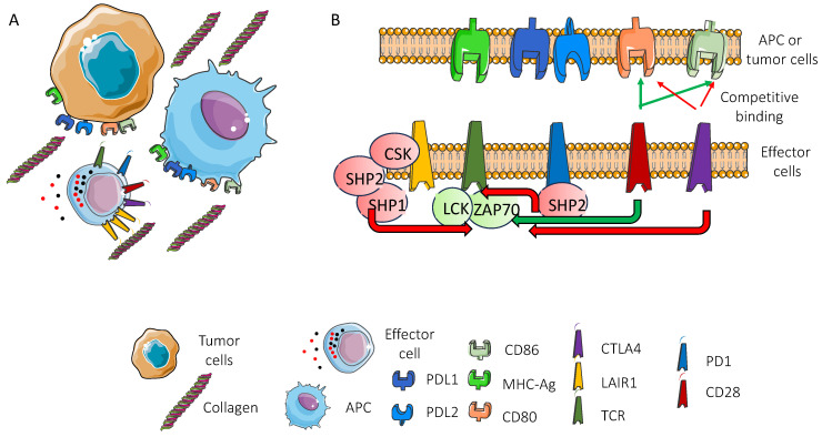
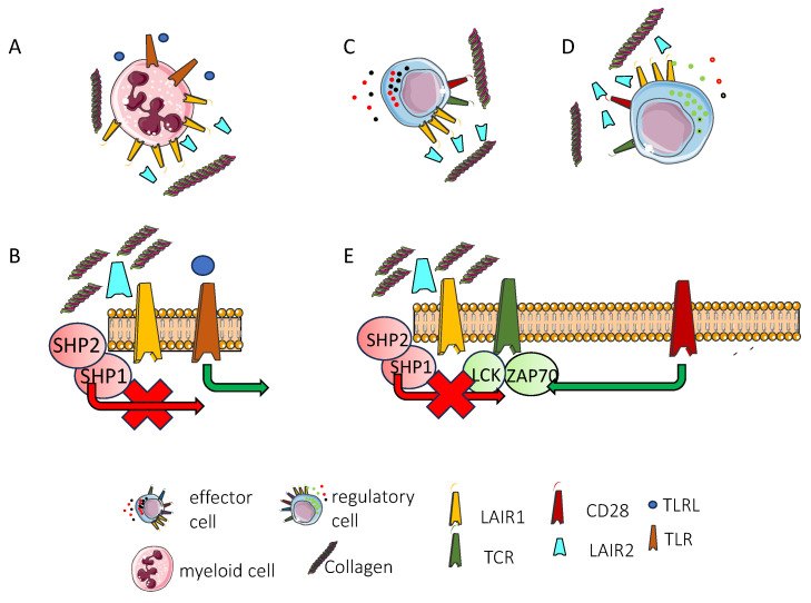
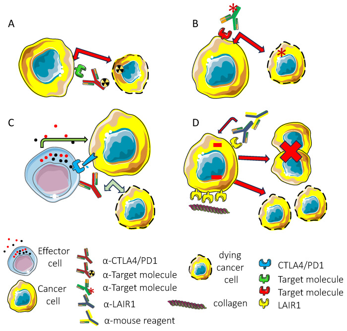
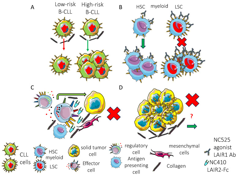
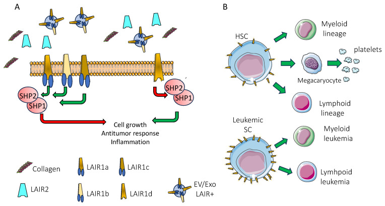
It is becoming evident that the therapeutic effect of reawakening the immune response is to limit tumor cell growth and expansion. The use of immune checkpoint inhibitors, like blocking antibodies against programmed cell death receptor (PD) 1 and/or cytotoxic T lymphocyte antigen (CTLA) 4 alone or in combination with other drugs, has led to unexpected positive results in some tumors but not all. Several other molecules inhibiting lymphocyte antitumor effector subsets have been discovered in the last 30 years. Herein, we focus on the leukocyte-associated immunoglobulin (Ig)-like receptor 1 (LAIR1/CD305). LAIR1 represents a typical immunoregulatory molecule expressed on almost all leukocytes, unlike other regulatory receptors expressed on discrete leukocyte subsets. It bears two immunoreceptor tyrosine-based inhibitory motifs (ITIMs) in the intracytoplasmic protein domain involved in the downregulation of signals mediated by activating receptors. LAIR1 binds to several ligands, such as collagen I and III, complement component 1Q, surfactant protein D, adiponectin, and repetitive interspersed families of polypeptides expressed by erythrocytes infected with Plasmodium malariae. This would suggest LAIR1 involvement in several cell-to-cell interactions and possibly in metabolic regulation. The presence of both cellular and soluble forms of LAIR would indicate a fine regulation of the immunoregulatory activity, as happens for the soluble/exosome-associated forms of PD1 and CTLA4 molecules. As a consequence, LAIR1 appears to play a role in some autoimmune diseases and the immune response against tumor cells. The finding of LAIR1 expression on hematological malignancies, but also on some solid tumors, could open a rationale for the targeting of this molecule to treat neoplasia, either alone or in combination with other therapeutic options.

Keywords: LAIR1; immune checkpoint; immune regulation; immunotherapy; inhibitory receptors.


Conflict of interest statement

The authors declare no conflicts of interest.

Figures

Figure 1
Figure 1
LAIR1-mediated molecular mechanisms of inhibition of immune cell function. (A) effector cells (NK or T lymphocytes) can interact with antigen-presenting cells (APCs, depicted in light blue) or tumor cells (brown cell) through different receptor–ligand pairs represented with different colors. Collagen, a main component of the extracellular matrix, is present among cells. LAIR1 (light brown receptor) is expressed on effector lymphocytes. As explained in the text, LAIR1 can deliver an inhibitory signal after interaction with collagen. The receptors expressed on lymphocytes such as TCR, CD28, PD1, and CTLA4 can deliver activating or inhibitory signals according to their association with transducer molecules. The expression of LAIR1 on APC or tumor cells is not shown in this figure to simplify the scenario. The figure is focused on TCR-mediated signal transduction in T lymphocytes. Here, the response of NK cells is not shown. (B) the interaction among the receptors on lymphocytes and ligands expressed on either APC or tumor cells has been magnified to show in detail the molecular interactions among the different molecules and the transducer molecules to which these receptors can be associated. The recognition of the antigen is mediated by the T cell receptor (depicted in dark green, first signal), which in turn activates the immune response. A second signal is delivered by the binding of CD28 (depicted in dark red) with CD80 or CD86 molecules. TCR and CD28 molecules concur in the activation of kinases such as LCK and/or ZAP70. The second co-stimulatory signal is needed to achieve the full activation of the effector cell, leading to lymphocyte proliferation, the killing of tumor target cells, or the production of cytokines. The regulatory receptor CTLA4 competes with CD28 for binding with CD80 or CD86, impairing the delivery of the second signal. PD1 interacting with PDL1 or PDL2 triggers the SHP2 phosphatase activity to block TCR signaling, impairing the activation of LCK and/or ZAP70 kinases. LAIR1 binds to collagen and associates with SHP1 or SHP2, similarly to PD1, which can reduce the phosphorylation of ZAP70 or LCK. Also, LAIR1 can engage the C-terminal Src kinase (CSK), regulating the receptor signal-mediated activation through tyrosine phosphorylation. The red lines indicate the inhibitory effect, while the green one indicates the activating effect. Templates of cell and molecule are from https://smart.servier.com/.
Figure 2
Figure 2
LAIR2 can regulate the signal delivered through LAIR1. (A) myeloid cells (pink) expressing the LAIR1 molecule (light brown) can be involved in inflammation processes, and LAIR2 (light blue), a soluble form of LAIR1, can be present in the microenvironment. LAIR2 has a greater affinity for collagen than LAIR1 and it can compete with LAIR1 for binding to collagen. The engagement of LAIR1 by collagen delivers an inhibitory signal. Competition with LAIR2 relieves the cells from this inhibitory signal. The net effect of this competition will depend on the level of expression of LAIR1, the concentration of LAIR2, and the presence of collagen in the microenvironment. (B) the collagen engagement of LAIR1 downregulates inflammation in myeloid cells. Indeed, the activating signal delivered through the Toll-like receptor (TLR) ligand (blue circle)–TLR (brown receptor) interaction is downregulated by SHP1/SHP2 phosphatases (red arrow). LAIR2, competing with LAIR1 for binding with collagen, blocks the negative signal (red cross) and allows the delivery of TLR-mediated activation (green arrow). (C) effector lymphocytes can be activated by recognition of the antigen. (D) regulatory cells inhibit several functions of effector cells by releasing immunomodulating cytokines. (E) the triggering of T cells through TCR (dark green) and CD28 (dark red) leading to the activation of LCK and ZAP70 is downregulated by LAIR1 binding with collagen (red arrow). LAIR2 inhibits (red cross) this signal by competition with LAIR1 allowing to TCR-mediated activation. It is conceivable that LAIR2 facilitates the activation of effector cells. Likewise, the inhibitory function of regulatory T cells can be elicited in the presence of LAIR2. Templates of cell and molecule are from https://smart.servier.com/.
Figure 3
Figure 3
Targeting of tumor cells with antibodies to tumor-expressed molecules. Antibodies target tumor surface molecules, leading to the inhibition of tumor cell growth and dissemination. The antibodies can recognize “target molecules” highly expressed on tumor cells (green and red receptors). The antibody-mediated effect can be elicited through different molecular mechanisms. (A) the antibody conjugated to a radioisotope recognizes the target molecule, and after internalization in tumor target cells, the radioisotope induces cell death. (B) the antibody linked to a cytotoxic drug (usually inhibitors of microtubule poly or depolymerization) (*) interacts with the target molecule; after internalization, the drug is released and it blocks essential biological components of the tumor target cell involved in cell proliferation and/or the maintenance of cellular homeostasis; (C) antibodies to classical immune checkpoint receptor molecules such as CTLA4 and/or PD1 can impair the binding of the inhibitory receptor expressed on lymphocytes with the corresponding ligand on tumor cells. As a consequence, the inhibitory receptor does not deliver an inhibitory signal on the activation of effector cell functions triggered through activating surface receptors such as TCR. This can allow effector T cells to exert their antitumor activity by recognizing the tumor antigens, releasing cytotoxic molecules or antitumor cytokines. The result of this effect is the killing of the tumor cell. (D) The inhibitory molecule expressed on tumor target cells (such as LAIR1 on leukemic cells) upon its cross-linking achieved by interaction with the ligand or appropriate second reagents can lead to an inhibitory signal in tumor cells. This signal can impair cell proliferation and/or induce cell death. It should be noted that the anti-LAIR1 mAb can function similarly to anti-PD1 when expressed on effector cells, while the engagement of LAIR1 on tumor cells, typically in hematological malignancies, can directly evoke inhibitory signals in tumor cells. Experimentally, the cross-linking of LAIR1 is achieved using an anti-mouse reagent. In vivo, this cross-linking is triggered by collagen, which expresses repetitive binding sites allowing the engagement of multiple LAIR1 molecules. Red arrows indicate negative signals, while the green ones indicate positive signals. The red cross indicates the block of proliferation. This figure does not show the effect of therapeutic antibodies mediated by immune cells such as antibody-dependent cellular cytotoxicity or complement dependent cytotoxicity. Templates of cell and molecule are from https://smart.servier.com/.
Figure 4
Figure 4
LAIR1 can play a role in the regulation of growth of some hematological malignancies, while in solid tumors its function is not well-defined. (A) LAIR1 expression levels and effects on CLLs. LAIR1 is expressed at different levels on the cell surface of B cells of chronic lymphocytic leukemia (CLL). Low-risk (LR) patients expressed high levels of LAIR1, conversely to high-risk (HR) patients. It is conceivable that LAIR1 engagement by collagen does not block the proliferation of HR CLLs (green arrow), while LR CLLs are controlled by LAIR1-mediated inhibitory signal (red arrow). (B) LAIR1 effects on myeloid leukemia. High levels of surface expression of LAIR1 on myeloid leukemia stem cells (LSCs) allow the delivery of an inhibitory signal through the LAIR1 agonist antibody (NC525) leading to LSC cell death and blocking leukemia proliferation (red cross). The NC525 antibody does not alter the growth of healthy hematopoietic stem cells (HSCs) because the LAIR1 level is too low to inhibit proliferation or induce cell death (green arrow). (C) therapeutic role of LAIR2. The LAIR2-Fc protein (NC410) can compete with surface LAIR1 for binding with collagen. This reduces/abolishes the LAIR1-mediated negative signal, allowing the activation of antitumor cells against tumor target cells. The net effect will depend on the presence of effector and/or regulatory cells in the tumor microenvironment expressing LAIR1. If effector cells prevail over regulatory cells, the tumor will not grow (red cross). (D) effects in solid tumors. LAIR1 expressed on solid tumors is associated with protumor or antitumor effects (see Table 3) leading to either tumor cell proliferation or tumor cell death. Deeper phenotypic and functional analyses are needed to properly define its role in solid tumor biology (red question mark). Templates of cell and molecule are from https://smart.servier.com/.
Figure 5
Figure 5
Focus on some of the possible studies to fill the gaps of knowledge on the function of LAIR1. (A) the expression and function of the LAIR1 isoforms (brown) can give some insights on the differential role of these molecules expressed on the several leukocytes subsets. It should be noted that the presence of the LAIR1d isoform (on the right) lacking the ITIM motifs can impair the signal mediated by the other LAIR isoforms bearing these ITIMs (on the left). The presence in the microenvironment of LAIR2 (light blue), collagen, and microvesicles/exosomes (EV/Exo LAIR) expressing isoforms of LAIR1 may directly influence the signal elicited upon the engagement of LAIR1 expressed at the cell surface. The binding of collagen with LAIR1a or LAIR1b or LAIR1c isoforms activates SHP1 and SHP2, blocking the function of the cells while binding with LAIR1d does not elicit the inhibitory signal, allowing cell growth, antitumor activities, and the triggering of inflammation. (B) a detailed analysis of the expression and effects of LAIR1 isoforms’ engagement on hematopoietic stem cells (HSCs) regulating differentiation of the myeloid and lymphoid lineages and platelets could help to better plan the targeting of leukemic stem cells (LSCs) to plan therapies to eliminate myeloid or lymphoid leukemias. Templates of cell and molecule are from https://smart.servier.com/.

Similar articles

References

    1. Zhang B., Liu J., Mo Y., Zhang K., Huang B., Shang D. CD8+ T cell exhaustion and its regulatory mechanisms in the tumor microenvironment: Key to the success of immunotherapy. Front. Immunol. 2024;15:1476904. doi: 10.3389/fimmu.2024.1476904. - DOI - PMC - PubMed
    1. Poggi A., Zocchi M.R. Natural killer cells and immune-checkpoint inhibitor therapy: Current knowledge and new challenges. Mol. Ther. Oncolytics. 2021;24:26–42. doi: 10.1016/j.omto.2021.11.016. - DOI - PMC - PubMed
    1. Jia H., Yang H., Xiong H., Luo K.Q. NK cell exhaustion in the tumor microenvironment. Front. Immunol. 2023;14:1303605. doi: 10.3389/fimmu.2023.1303605. - DOI - PMC - PubMed
    1. Bald T., Krummel M.F., Smyth M.J., Barry K.C. The NK cell-cancer cycle: Advances and new challenges in NK cell-based immunotherapies. Nat. Immunol. 2020;21:835–847. doi: 10.1038/s41590-020-0728-z. - DOI - PMC - PubMed
    1. Baessler A., Vignali D.A.A. T Cell Exhaustion. Annu. Rev. Immunol. 2024;42:179–206. doi: 10.1146/annurev-immunol-090222-110914. - DOI - PubMed

Substances

Grants and funding

LinkOut - more resources