. 2025 Jun 16;15(6):873.
doi: 10.3390/biom15060873.

Targeting Cellular Senescence to Enhance Human Endometrial Stromal Cell Decidualization and Inhibit Their Migration

Affiliations

Targeting Cellular Senescence to Enhance Human Endometrial Stromal Cell Decidualization and Inhibit Their Migration

Julia Delenko et al. Biomolecules. .

Abstract

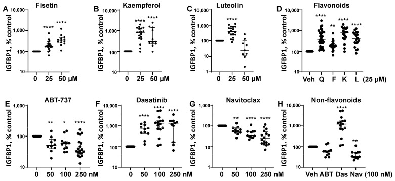
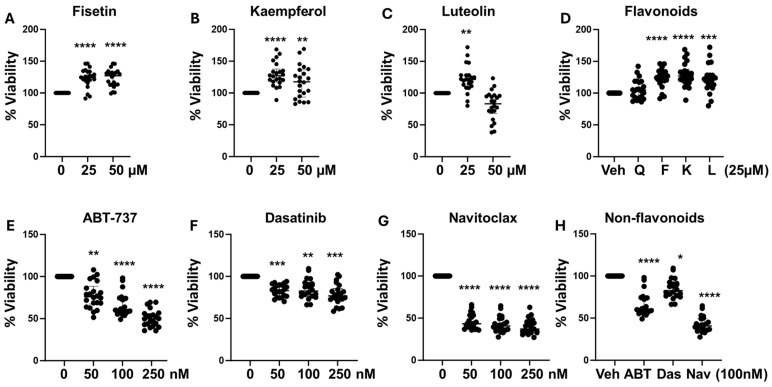
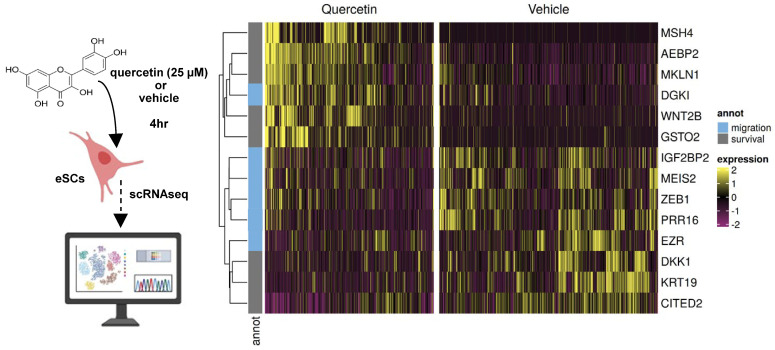
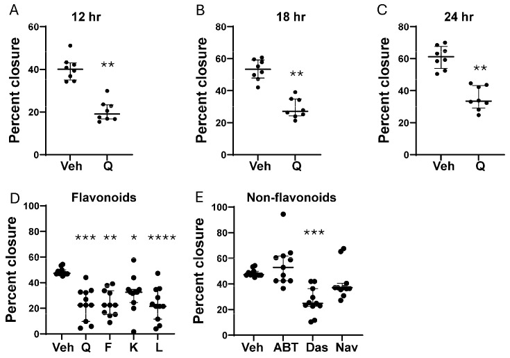
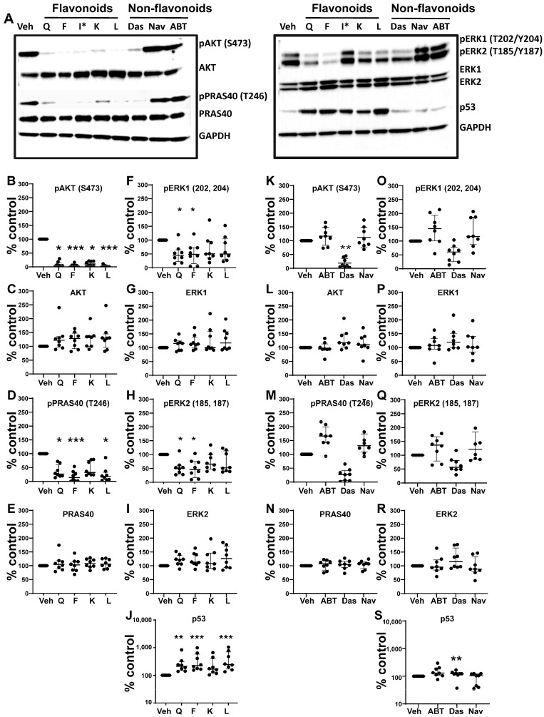
Cellular senescence leads to stable cell cycle arrest and an inflammatory senescence-associated secretory phenotype that varies with stressor and cell type. To mitigate these effects and improve health, senotherapeutics (e.g., senolytics and senomorphics) have been developed. Senescent-like endometrial stromal cells (eSCs) lining the uterus of patients with endometriosis and infertility are proposed to impair decidualization, a differentiation process required for uterine receptivity in humans. Quercetin, a natural flavonoid senolytic, dramatically improves decidualization and reduces endometriosis in rodent models. However, little is known about the comparative effects of various senotherapeutics on eSCs. Using menstrual effluent-derived eSCs, we evaluated the effects of flavonoid and non-flavonoid compounds on eSC functions associated with endometriosis, aiming to identify optimal senotherapeutics for future clinical trials. Among flavonoids tested, all senolytics (quercetin, fisetin, and luteolin) and kaempferol, a senomorphic, significantly improved decidualization without cytotoxicity. Although non-flavonoids exhibited notable cytotoxicity, dasatinib, but neither ABT-737 nor navitoclax, enhanced decidualization. Flavonoid senotherapeutics and dasatinib significantly inhibited eSC migration. Mechanistic studies revealed that all flavonoids and dasatinib suppressed AKT phosphorylation and upregulated p53 expression. Notably, only quercetin and fisetin reduced ERK1/2 phosphorylation. Furthermore, flavonoid-senolytics and dasatinib consistently eliminated senescent eSCs. These findings support future studies to assess the therapeutic potential of in vivo supplementation with flavonoid senolytics on eSC function using menstrual effluent.

Keywords: endometriosis; flavonoids; infertility; menstrual effluent; quercetin; senescence; senolytic; senomorphic.


Conflict of interest statement

A.G.B., L.F. and D.M.E. are co-founders of Senesys Bio, delivering a new generation of targeted senolytics.

Figures

Figure 1
Figure 1
Flavonoid and non-flavonoid senotherapeutics differentially affect human endometrial stromal cell (eSC) decidualization. (AD) Human eSCs were treated with flavonoids: fisetin (A), kaempferol (B), and luteolin (C) at 0–50 µM (0 = vehicle) or (D) all flavonoids (quercetin, Q; fisetin, F; kaempferol, K; luteolin, L) at 25 µM vs. vehicle (Veh) for 4 h prior to cAMP + MPA. Decidualization was determined by measuring IGFBP1 production in the culture supernatants 48 h later by ELISA. (EH) eSCs were treated with non-flavonoids: ABT-737 (E), dasatinib (F), and navitoclax (G) at 0–250 nM (0 = vehicle) or all non-flavonoids (ABT-737, ABT; dasatinib, Das; navitoclax, Nav) at 100 nM vs. vehicle (Veh) (H) for 4 h prior to decidualization and assessment of IGFBP1, as described above. Data are shown % control IGFBP1 values with vehicle-treated cells = 100%. Each point represents mean data from one individual’s eSCs, with median and IQR shown for each group. * p < 0.05 vs. Veh; ** p < 0.01 vs. Veh; **** p < 0.0001 vs. Veh.
Figure 2
Figure 2
Flavonoid senotherapeutics are mostly non-cytotoxic compared to non-flavonoid senotherapeutics. (AD) Human eSCs were treated with flavonoids: fisetin (A), kaempferol (B), and luteolin (C) at 0–50 µM (0 = vehicle) or (D) all flavonoids at 25 µM (quercetin, Q; fisetin, F; kaempferol, K; luteolin, L) vs. vehicle (Veh) for 48 h and assessed for cytotoxicity using the neutral red assay. (EH) Human eSCs were treated with non-flavonoids, ABT-737 (E), dasatinib (F), and navitoclax (G) at 0–250 nM (0 = vehicle) or all non-flavonoids at 100 nM (H) (ABT-737, ABT; dasatinib (Das); navitoclax (Nav) vs. vehicle (Veh) and assessed for cytotoxicity by neutral red. Data are shown as % control viability values with vehicle-treated cells = 100%. Each point represents mean data from one individual’s eSCs, with median and IQR shown for each group.* p < 0.05, ** p < 0.01 vs. Veh; *** p < 0.001 vs. Veh **** p < 0.0001 vs. Veh.
Figure 3
Figure 3
Human eSCs (n = 4 healthy controls) were treated with quercetin (25 µM) vs. vehicle for 4 h and then analyzed by scRNAseq. Heat map shows differentially expressed genes implicated in cell migration and survival.
Figure 4
Figure 4
The effects of flavonoid and non-flavonoid senotherapeutics on eSC migration. (AC) Confluent eSCs were treated with vehicle (Veh) or quercetin (Q) at 25 µM and then analyzed for migration using the wound closure assay 12 h (A), 18 h (B), and 24 h (C) post-scratch (n = 8 subjects). Cell migration was assessed as percentage wound closure at each time point. (D) Confluent eSCs (n = 11 subjects) were treated with flavonoids: quercetin (Q), fisetin (F), kaempferol (K), and luteolin (L) at 25 µM vs. vehicle (Veh). (E) eSCs (n = 11 subjects) were treated with vehicle or non-flavonoids (ABT-737 (ABT), dasatinib (Das), navitoclax (Nav)) at 100 nM and wound closure was compared to vehicle (Veh)-treated cells. (D,E) Data are shown as percentage closure measured at the time when vehicle-treated cells showed approximately 50% closure. Each point represents mean data from one individual’s eSCs, with median with IQR shown for each group. * p < 0.05 vs. Veh; ** p < 0.01 vs. Veh; *** p < 0.001 vs. Veh **** p < 0.0001 vs. Veh.
Figure 5
Figure 5
The differential effects of flavonoid and non-flavonoid senotherapeutics on eSC signaling pathways. (A) Representative western blot images for one control participant’s eSCs treated with vehicle (Veh) vs. senotherapeutics, flavonoids: quercetin (Q), fisetin (F), kaempferol (K), and luteolin (L) at 25 µM and non-flavonoids: ABT-737 (ABT), dasatinib (Das), navitoclax (Nav)) at 100 nM. Note: I* was not included in the analyses. (BJ) eSCs (n = 8 subjects) were treated with vehicle (Veh) or flavonoids, including quercetin (Q), fisetin (F), kaempferol (K), or luteolin (L) at 25 µM for 4 h before analyzing cell lysates by western blotting for p-AKT (B), total AKT (C), p-PRAS40 (D), total PRAS40 (E), p-ERK1 (F), total ERK1 (G), p-ERK2 (H), total ERK2 (I), and p53 (J). (KS) eSCs (from the same 8 subjects) were treated with vehicle (Veh) or various flavonoids, including ABT-737 (ABT), dasatinib (Das), or navitoclax (Nav) for 4 h before analyzing cell lysates for the same proteins as above. Band densities were normalized to GAPDH and corrected for total non-phosphorylated proteins, where appropriate, and shown as % control between Veh- vs. flavonoid-treated eSCs (BJ) or Veh- vs. non-flavonoids (KS), with vehicle-treated cell values set to 100%. Each point represents mean data from one individual’s eSCs (from 8 subjects), with median and IQR shown for each group. * p < 0.05; ** p < 0.01; *** p < 0.001. All uncropped blot images and band densities (with calculations) are shown in Supplemental Data S1 and S2, respectively.
Figure 6
Figure 6
Senotherapeutics effectively eliminate senescent cells. (A,B) Vehicle-eSCs vs. palbociclib (PALB)-treated eSCs (to induce senescence) analyzed for NanoJagg (NJ) accumulation (n = 10 subjects), with data shown as NJ uptake per cell as mean paired data connected by a line for each participants’ eSCs (A) and NJ uptake per cell as fold-change, where vehicle-treated eSCs = 1; each point represents mean data from one individual’s eSCs, with median and IQR shown for each group (B). (C,D) Vehicle-treated eSCs (C) or PALB-treated (senescent) eSCs (D) treated with vehicle or quercetin (at 10 or 25 µM) and assessed for NJ uptake (3 subjects each). Data are shown as mean paired data connected by a line. (E) Western blot showing NJ−positive (NJ+) vs. NJ−negative (NJ-) sorted eSCs (lanes 1–5) and non-sorted eSCs ± quercetin (Q, 25 µM) (lanes 6–9). Lanes 1–5: paired NJ+ vs. NJ following vehicle-(V) or palbociclib (PL)-treated eSCs from subject 1 (V1 and PL1), subject 2 (V2 and PL2) and subject 3 (PL3) blotted with antibodies to senescence markers (p-Rb and p16) and GAPDH. Note V2 eSCs are low passage (p2) and other eSCs are p6–p8. Lanes 6–9: eSCs from subject 4 (p6–p8) were pre-treated with vehicle (V4) vs. PALB (PL4) to induce senescence followed by vehicle (Veh) vs. quercetin (Q, 25 µM) and then blotted with antibodies to senescence markers (p-Rb and p16) and GAPDH. Full blots and band densities are in Supplemental Data S5. (F,G) Comparison of vehicle-eSCs vs. PALB-eSCs for cell proliferation (n = 8 subjects); F shows paired data (relative cell number) connected by a line for each participants’ eSCs and G shows proliferation data (relative cell number) where each point represents mean data from one individual’s eSCs, with median with IQR shown for each group. (HK) eSCs (from 11 subjects) were either pre-treated with vehicle (Veh) (H,J) or PALB (I,K) to induce senescence before treatment with flavonoids (H,I) or non-flavonoids (J,K) and then assessed for NJ accumulation (shown as NJ uptake per cell as fold-change as in (B)). Each point represents mean data from one individual’s eSCs, with median and IQR shown for each group. * p < 0.05; ** p < 0.01; *** p  <  0.001; **** p < 0.0001.

Similar articles

References

    1. Kirkland J.L., Tchkonia T. Cellular Senescence: A Translational Perspective. EBioMedicine. 2017;21:21–28. doi: 10.1016/j.ebiom.2017.04.013. - DOI - PMC - PubMed
    1. Gorgoulis V., Adams P.D., Alimonti A., Bennett D.C., Bischof O., Bishop C., Campisi J., Collado M., Evangelou K., Ferbeyre G., et al. Cellular Senescence: Defining a Path Forward. Cell. 2019;179:813–827. doi: 10.1016/j.cell.2019.10.005. - DOI - PubMed
    1. Reyes A., Ortiz G., Duarte L.F., Fernandez C., Hernandez-Armengol R., Palacios P.A., Prado Y., Andrade C.A., Rodriguez-Guilarte L., Kalergis A.M., et al. Contribution of viral and bacterial infections to senescence and immunosenescence. Front. Cell Infect. Microbiol. 2023;13:1229098. doi: 10.3389/fcimb.2023.1229098. - DOI - PMC - PubMed
    1. L’Hote V., Mann C., Thuret J.Y. From the divergence of senescent cell fates to mechanisms and selectivity of senolytic drugs. Open Biol. 2022;12:220171. doi: 10.1098/rsob.220171. - DOI - PMC - PubMed
    1. Lelarge V., Capelle R., Oger F., Mathieu T., Le Calve B. Senolytics: From pharmacological inhibitors to immunotherapies, a promising future for patients’ treatment. NPJ Aging. 2024;10:12. doi: 10.1038/s41514-024-00138-4. - DOI - PMC - PubMed

LinkOut - more resources