. 2025 Jun 17;15(6):883.
doi: 10.3390/biom15060883.

Pre-Amplification of Cell-Free DNA: Balancing Amplification Errors with Enhanced Sensitivity

Affiliations

Pre-Amplification of Cell-Free DNA: Balancing Amplification Errors with Enhanced Sensitivity

Wei Yen Chan et al. Biomolecules. .

Abstract

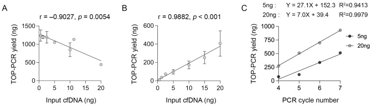
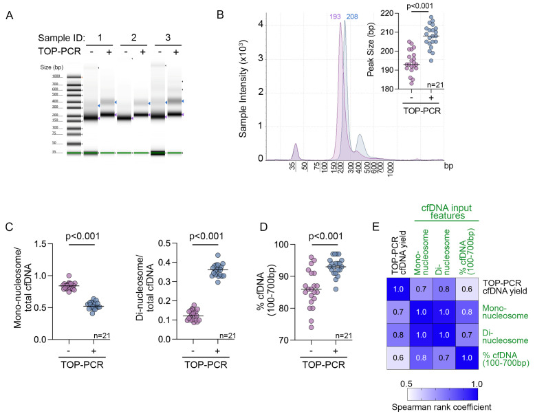
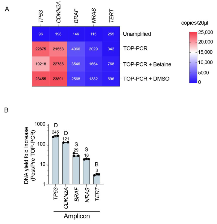
Circulating tumour DNA (ctDNA) is a promising biomarker for personalised oncology. However, its clinical utility is limited by detection sensitivity, particularly in early-stage disease. T-Oligo Primed Polymerase Chain Reaction (TOP-PCR) is a commercial amplification approach utilising an efficient "half-adapter" ligation design and a single-primer-based PCR strategy. This study evaluated the clinical value and application of cell-free DNA (cfDNA) pre-amplification. cfDNA amplification with TOP-PCR preserved DNA size profiles and resulted in a 22 bp size increase due to the half-adaptor ligation. Gene target amplification rates varied, showing lower efficiency for the GC-rich TERT promoter amplicon and higher efficiency for the BRAF and TP53 amplicons. Optimised pre-amplification (20 ng cfDNA input and 5-7 cycles of PCR) enhanced ctDNA detection sensitivity and expanded sample availability for the detection of multiple tumour-informed mutations. Importantly, PCR errors emerged in pre-amplified cfDNA samples, underscoring the necessity for negative controls and the establishment of stringent mutation positivity thresholds.

Keywords: circulating tumour DNA; detection sensitivity; melanoma.


Conflict of interest statement

J.H.L. received honorarium from MSD, BMS, Sanofi, Roche, AstraZeneca, and Novartis and sits of advisory boards for MSD, BMS, and Sanofi. R.A.S. has received fees for professional services from SkylineDx BV, IO Biotech ApS, MetaOptima Technology Inc., F. Hoffmann-La Roche Ltd., Evaxion, Provectus Biopharmaceuticals Australia, Qbiotics, Novartis, Merck Sharp & Dohme, NeraCare, AMGEN Inc., Bristol-Myers Squibb, Myriad Genetics, and GlaxoSmithKline. G.V.L. is a consultant advisor for Agenus, Amgen, Array Biopharma, AstraZeneca, Bayer, BioNTech, Boehringer Ingelheim, Bristol Myers Squibb, Evaxion, Hexal AG (Sandoz Company), Highlight Therapeutics S.L., IOBiotech, Immunocore, Innovent Biologics USA, Merck Sharpe & Dohme, Novartis, PHMR Ltd., Pierre Fabre, Regeneron, Scancell, and SkylineDX B.V. All remaining authors have declared no conflicts of interest.

Figures

Figure 1
Figure 1
The performance of TOP-PCR. The total cfDNA yield post-TOP-PCR amplification is shown relative to the initial cfDNA PCR input after (A) 15 cycles of amplification (at least two independent amplifications per input cfDNA) and (B) 5 cycles of amplification (n = 1 for <5 ng cfDNA input, at least two independent amplifications for all other input cfDNA amounts). Pearson correlation coefficient and two-tailed p-values are shown for each graph. (C) Amplification efficiency was tested using 5 ng and 20 ng inputs and four to seven PCR cycles (n = 1 amplification reaction from a dilution series of a single cfDNA sample mix). The regression line equations and goodness-of-fit data (R2) are also shown. Data shown are mean ± s.d; ng, nanogram.
Figure 2
Figure 2
The effect of TOP-PCR on cfDNA mono-nucleosome and di-nucleosome peaks. (A) The electrophoresis of melanoma cfDNA pre (-) and post (+) TOP-PCR amplification (20 ng cfDNA input, five cycles of amplification). cfDNA derived from three melanoma patients was separated using the Agilent TapeStation 4150. Purple arrows indicate the mono-nucleosomal DNA, and blue arrows indicate the di-nucleosomal DNA (where visible). The molecular weight ladder (shown on the left) includes two internal standards (lower and upper markers); variation in their appearance across samples is due to image scaling adjustments made for visual display. The TapesStation 4150 electrophoresis without intensity scaling is shown in Figure S1. (B) The size profile of representative patient-matched cfDNA pre- and post-TOP-PCR amplification. (Inset) The scatter plot showing the median mono-nucleosome peak size for 21 stage IV melanoma patient-matched cfDNA samples pre- and post-TOP-PCR amplification (20 ng cfDNA input, seven PCR cycles). Median is shown in each graph. Peak size data were compared using Wilcoxon matched-pairs signed rank test. (C) The yield (quantitated on TapeStation 4150) of mono-nucleosome (left) and di-nucleosomal (right) cfDNA pre- and post-TOP-PCR amplification of 20 ng cfDNA input for seven PCR cycles is shown relative to the total cfDNA yield (100–700 bp) in 21 stage IV melanoma patient-matched cfDNA samples. Median is shown in each graph. Data were compared using the Wilcoxon matched-pairs signed-rank test. (D) The percentage of DNA between 100 and 700 bp (quantitated on TapeStation 4150) is shown pre- and post-TOP-PCR amplification (20 ng cfDNA input, seven PCR cycles) in 21 stage IV melanoma patient-matched samples. Median is shown in each graph. Data were compared using the Wilcoxon matched-pairs signed-rank test. (E) The correlation matrix of DNA yield following TOP-PCR amplification (20 ng input, seven PCR cycles) with cfDNA features including cfDNA mono-nucleosome input, cfDNA di-nucleosome input, and % cfDNA input (100–700 bp DNA fraction). Data derived from 21 stage IV melanoma patients. The Spearman rank correlation coefficients are shown within the matrix. Correlation p-values are all <0.01.
Figure 3
Figure 3
Gene-specific effects on pre-amplification efficacy. (A) Heat map showing the yield (copies/20 µL in ddPCR) of indicated gene targets without pre-amplification, or after 15 cycles of TOP-PCR amplification with no molecular enhancers (TOP-PCR) or including 0.5 M betaine or 5% DMSO. The mean DNA yield of 2–3 independent PCR reactions is shown in each cell. Template cfDNA was derived from melanoma cell line culture media. The CDKN2A assays were performed using cfDNA from HDF1314-derived supernatant, while the remaining four assays were conducted from purified DNA from melanoma cell lines. (B) The maximum fold increase in DNA yield (copies/20 µL; post/pre-TOP-PCR) is shown for each target amplicon after 15 cycles of TOP-PCR amplification. This value represents the highest amplification yield observed for each target, highlighting the most effective enhancer (above each column) for each amplicon. The corresponding optimum amplification conditions for each DNA target is indicated: S—standard TOP-PCR conditions with no enhancer; B—TOP-PCR with 0.5 M betaine; D—TOP-PCR with 5% DMSO.

Similar articles

References

    1. Peng Y., Mei W., Ma K., Zeng C. Circulating Tumor DNA and Minimal Residual Disease (MRD) in Solid Tumors: Current Horizons and Future Perspectives. Front. Oncol. 2021;11:763790. doi: 10.3389/fonc.2021.763790. - DOI - PMC - PubMed
    1. Lee J.H., Long G.V., Boyd S., Lo S., Menzies A.M., Tembe V., Guminski A., Jakrot V., Scolyer R.A., Mann G.J., et al. Circulating tumour DNA predicts response to anti-PD1 antibodies in metastatic melanoma. Ann. Oncol. 2017;28:1130–1136. doi: 10.1093/annonc/mdx026. - DOI - PubMed
    1. Lee J.H., Saw R.P., Thompson J.F., Lo S., Spillane A.J., Shannon K.F., Stretch J.R., Howle J., Menzies A.M., Carlino M.S., et al. Pre-operative ctDNA predicts survival in high-risk stage III cutaneous melanoma patients. Ann. Oncol. 2019;30:815–822. doi: 10.1093/annonc/mdz075. - DOI - PMC - PubMed
    1. Ignatiadis M., Sledge G.W., Jeffrey S.S. Liquid biopsy enters the clinic-implementation issues and future challenges. Nat. Rev. Clin. Oncol. 2021;18:297–312. doi: 10.1038/s41571-020-00457-x. - DOI - PubMed
    1. Chin R.I., Chen K., Usmani A., Chua C., Harris P.K., Binkley M.S., Azad T.D., Dudley J.C., Chaudhuri A.A. Detection of Solid Tumor Molecular Residual Disease (MRD) Using Circulating Tumor DNA (ctDNA) Mol. Diagn. Ther. 2019;23:311–331. doi: 10.1007/s40291-019-00390-5. - DOI - PMC - PubMed

MeSH terms

LinkOut - more resources