Review
. 2025 Jun 9;17(12):1915.
doi: 10.3390/cancers17121915.

Cryoablation and Intratumoral Immunotherapy for Breast Cancer: A Future Path to Cost-Effective De-Escalation for Larger Tumors, Lymph Nodes and Metastatic Disease

Affiliations
Review

Cryoablation and Intratumoral Immunotherapy for Breast Cancer: A Future Path to Cost-Effective De-Escalation for Larger Tumors, Lymph Nodes and Metastatic Disease

Josephine Fermanian et al. Cancers (Basel). .

Abstract

Cryoablation is a promising, cost-effective option to de-escalate surgical breast cancer morbidity, but presently is only suggested for breast cancers < 1.5 cm, in select candidates. Breast cancer cryoablation is not a reliably covered procedure by insurance and is mainly guided by ultrasound (US), using a single cryoprobe. Yet, cryoablation is an accepted treatment option for various malignancies, including those of the kidney, liver and lung, utilizing a predominantly CT-guided, multi-probe approach using crucial cytotoxic isotherms for thorough tumor coverage. Cryoablation thus continues to find new clinical utility and is rapidly advancing on multiple fronts, similar to immunotherapy. Clinical concerns of expanding cryoablation to breast tumors > 1.5 cm is more related to the greater risk of metastatic spread to local lymph nodes and beyond. Combined adjuvant treatment, such as radiation and/or chemotherapy, are currently used for regional and systemic breast cancer control, but have significant associated morbidities. US/CT-guided multi-probe large-volume breast cryoablation is presented as a thorough local control option for select patients. Intratumoral chemotherapy by direct tumor injection has been shown to be safe and is currently being tested with immunotherapy drugs and exhibits much lower morbidity. Cryoablation combined with intratumoral immunotherapy is presented to show robust systemic immune response and the potential to provide additional protection from regional and/or metastatic disease spread while de-escalating the morbidities from current adjuvant treatments for larger breast cancers. While further clinical trials are needed, it is essential to pursue safe and effective breast cancer treatments that offer the potential for cost-efficiency and therapeutic de-escalation across a wide spectrum of breast cancer cases.

Keywords: breast cancer; cryoablation; de-escalation; immunotherapy; intratumoral.


Conflict of interest statement

P.J.L. acknowledges that he served as Chief Medical Officer of Rampart Health, LLC and is a co-author of the abstract presented at the 2023 SITC conference referenced in this review. He also discloses his current role as Chief Medical Consultant for Delphinus Medical Technologies, Inc., the manufacturer of the SoftVue device. All other authors declare no conflicts of interest.

Figures

Figure 1
Figure 1
The 1–2 rule for cryoprobe placements according to tumor size, whereby standard cryoprobes (2.1/2.4 mm diameter) are placed no farther than 1 cm from the tumor margin and <2 cm apart. If a single probe is used for very small masses, a precise central location is needed to cover the tumor with ice (outer gray circles around each probe), and lethal ice (smaller dashed line ~5 mm inside ice). Conversely, when more probes are used, the sum of the individual ice projections (darker gray circles) synergistically expand, extending lethal ice (larger dashed line) ~5 mm beyond all tumor margins while visible ice (lighter gray circle) projects ~10 mm.
Figure 2
Figure 2
Ref. [43] (a) Diagram shows basic isotherms for single (left and center) and double cryoprobes (right). Accurate central placement of a single 2.4 mm cryoprobe (left image, larger black arrow) within a simulated 1.5 × 1.5 cm tumor (dark gray) may still not produce sufficient lethal ice to cover all tumor margins (dashed line ~<30 °C, diameter ~1.5 cm). Even though visible ice (solid outer circle) may appear to cover all tumor margins, slight off-center placement (middle image, curved black arrow) leaves a grossly untreated tumor (red bracket) beyond the lethal isotherm (dashed circle). Tumor on the right is covered by lethal ice due to synergy produced by two cryoprobes [47]. (b) Avoiding posterior positive margins: heat load effects of the chest wall. The estimated temperature difference between the skin surface (30 °C) and chest wall/body (36 °C) causes greater heat load along the posterior margin of ice propagation, which narrows the posterior distance between the visible (0 °C) and lethal (–30 °C) isotherms (curved solid arrows). Ablation on the left shows the central position of cryoprobes and greater anterior extension of visible ice beyond the tumor margin; however, incomplete coverage of posterior tumor margins (red curved arrows) is noted, like that seen in prior series [40]. Ablation on the right shows thorough tumor coverage by lethal ice due to the more posterior placement of cryoprobes in the tumor (red straight arrows), thus overcoming the heat-sink effect along the chest wall [43].
Figure 3
Figure 3
Ref. [47]. Progression of the lethal isotherm (−30 °C) at 5 min (inner dashed white lines), 10 min (dashed intermediate lines), and 15 min (dashed outer lines) is shown for double and triple JT Ar-based 1.7 and 2.4 mm cryoprobes (larger central solid circles), overlaid on the CT image for the total ice appearance at 15 min. (a) With a double cryoprobe (largest 2 central bright circles) configuration, the lethal ice surface area grows more for 2.4 mm cryoprobes (right) after 5 min as a result of early synergy than for 1.7 mm cryoprobes (left). Smaller peripheral bright solid circles represent thermocouples documenting the isotherm temperatures. (b) With a triple-cryoprobe configuration, the lethal ice surface area grows more for 2.4 mm cryoprobes (right) after 5 minutes as a result of early synergy, but the difference becomes less over time after synergy also occurs for 1.7 mm cryoprobes (left). Smaller bright peripheral circles are again the temperature-validating thermocouples.
Figure 4
Figure 4
Hydrodissection planning and two components of the Knuckle Rule. For tumors <~5 mm from the surface, mobility should feel like the skin sliding over a knuckle (left) to allow the injected fluid to thicken the skin by >2 mm. If you can pick up the skin even a little (right), then you can thicken the overlying skin > 10 mm which is especially needed for large-volume ablations.
Figure 5
Figure 5
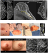
Ref. [43] (ad) Pre-cryoablation mammograms (a,b)-left (dashed circle)) show a large grouping of malignant calcifications, compatible with the CT enhancement (c,d) extending up to 3.4 cm in the lower lateral left breast on axial (c) and sagittal (d) images. Procedural images (eh) show initial planning marks on the skin (e) with thorough hydrodissection over the lateral aspect of the enhancing tumor (f). Five cryoprobes were placed medially (g) and extended throughout the interior breast as noted on the immediate postprocedural CT (h) showing a low-density ice coverage of ~8 cm craniocaudal. (il) The two post-procedure left images (i,j) show minimal residual bruising at day 15, with eventual resorption and mild overlying skin retraction of the left lower lateral breast by day 111 (~4 mo). The two right magnified mammograms compare the pre-cryoablation and 12-month follow-up appearance of the near complete resorption of the diffuse region of calcifications. The residual posterior calcifications appeared to be relatively stable and may represent residual benign calcifications but further long-term follow-up is needed to assess whether malignant calcifications resorb after cryoablation more so than benign calcifications. Moreover, no significant coarse calcifications of fat necrosis were noted. The described large volume breast cryoablation procedure required sufficient procedural experience and/or image guidance skills with US and CT, following careful consideration of the breast MR “roadmap”, and associated three-dimensional CT to plan for thorough coverage. However, our objective of ablating nearly any size breast tumor is also similar to multi-probe US/CT-guided procedures in nearly any other organ. For instance, Wang et al. found successful local control across a wide range of thoracic tumor sizes, highlighting the adaptability of cryoablation beyond strict size constraints. Similarly, Littrup et al. demonstrated that renal tumors of various sizes, including those >3 cm, were effectively managed through percutaneous cryoablation guided by CT. Moreover, in their long-term study of hepatic tumors, they showed that cryoablation provided consistent tumor control across a diverse patient cohort, including those with larger and anatomically complex lesions [48,51,52,53]. Holmes and Iyengar maintain that although cryoablation technology is optimized in ultrasound-visible stage 1 breast cancer, technique modifications can permit the cryoablation of stage 0, II, III and IV. The authors reveal that cryoablation achieves durable local tumor control in appropriately selected breast cancer patients with ongoing trials supporting its use as a definitive treatment modality in select populations. The authors emphasize that when combined with multidisciplinary oversight, cryoablation can serve as a viable alternative to surgery, offering effective control with minimal morbidity [10]. In summary, cryoablation of nearly any size breast cancer appears to be feasible, yet also may still require adjunctive treatments similar to surgical resection in order to manage the greater likelihood of nodal and distant metastatic disease from larger breast cancers. While current adjunctive treatments—such as hormonal therapy, radiation, and chemotherapy—are essential for managing regional and metastatic breast cancer, they are often associated with substantial side effects and morbidity. Similarly, intravenous immunotherapy carries a high risk of adverse effects. The likelihood of breast cancer recurrence after cryoablation is closely tied to ensuring complete cytotoxic coverage that extends beyond all visible tumor boundaries. Because of this, precise visualization of tumor margins may be even more critical in cryoablation than in surgical resection. Any residual viable tumor left behind may be stimulated by the surrounding hypervascular healing zone, potentially leading to rapid regrowth. Although this risk also exists with incomplete surgical excision, residual tumor proliferation following cryoablation can occur more aggressively, necessitating prompt re-treatment. The remaining sections in this review also offer hope that these recurrence risks, side effects and morbidities may also be diminished when cryoablation is combined with intratumoral immunotherapy. The dashed circles in figures (a,b,d) is the grouping of malignant calcifications.
Figure 6
Figure 6
Points to consider when designing an intratumoral immunotherapy clinical trial [64,65].
Figure 7
Figure 7
Top (AC) and bottom (A*–C*) row of images show 3 pre- and post-ablation immunotherapy comparisons across anatomic areas. In Pre-Cryo-Immunotherapy CT images, the 2 lung lesions are visualized in the right upper lobe (A) and right perihilar region (B), each measuring up to ~2 cm. Axillary lymph node metastasis is shown in (C), measuring up to 3.6 cm in maximum diameter. Center row (DF) display the ultrasound-guided cryoablation mid-procedure images confirming accurate cryoprobe insertion and iceball formation. Arrows in (E) show the single cryoprobe (blue arrow) and injection needle in (yellow) positioned ~7 mm from the cryoprobe. Image (F) (middle row, right) shows the 2 cm diameter echogenic ice rim with shadowing nearly covering all aspects of the tumor at 4-min freeze.  Bottom row of CT images (A*–C*) obtained 6-months post-cryo-immunotherapy show the same anatomic sites with substantial tumor reduction. Right lung lesions (A*, B*) show radiological resolution with no visible mass. In image (C*) axillary nodes shrinks to 14.7 × 25 mm, reflecting significant immunologic and ablative response.

References

    1. Surveillance, Epidemiology, and End Results (SEER) Program. Cancer Statistics for Breast Cancer. National Cancer Institute. [(accessed on 18 October 2024)]; Available online: https://seer.cancer.gov/statfacts/html/breast.html.
    1. Holmes D.R. Breast cancer care during a pandemic: An opportune time for cryoablation? Breast Cancer Res. Treat. 2020;182:515–521. doi: 10.1007/s10549-020-05724-0. - DOI - PMC - PubMed
    1. Sacchini V., Norton L. Escalating de-escalation in breast cancer treatment. Breast Cancer Res. Treat. 2022;195:85–90. doi: 10.1007/s10549-022-06685-2. - DOI - PubMed
    1. Khan S.Y., Cole J., Habrawi Z., Melkus M.W., Layeequr Rahman R. Cryoablation Allows the Ultimate De-escalation of Surgical Therapy for Select Breast Cancer Patients. Ann. Surg. Oncol. 2023;30:8398–8403. doi: 10.1245/s10434-023-14332-3. - DOI - PMC - PubMed
    1. Huang M.L., Tomkovich K., Lane D.L., Katta R., Candelaria R.P., Santiago L. Breast Cancer Cryoablation Fundamentals Past and Present: Technique Optimization and Imaging Pearls. Acad. Radiol. 2023;30:2383–2395. doi: 10.1016/j.acra.2023.05.019. - DOI - PMC - PubMed

LinkOut - more resources