. 2025 Jun 9;17(12):1920.
doi: 10.3390/cancers17121920.

The RXR Agonist MSU-42011 Reduces Tumor Burden in a Murine Preclinical NF1-Deficient Model

Affiliations

The RXR Agonist MSU-42011 Reduces Tumor Burden in a Murine Preclinical NF1-Deficient Model

Pei-Yu Hung et al. Cancers (Basel). .

Abstract

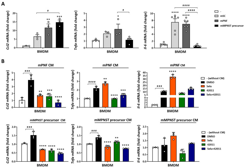
Background/Objectives: Neurofibromatosis type 1 (NF1) is a prevalent inherited disorder, with approximately 50% of affected individuals developing plexiform neurofibromas (PNFs), which can progress to highly aggressive malignant peripheral nerve sheath tumors (MPNSTs). While selumetinib is FDA-approved for PNFs, its efficacy in MPNSTs is limited and associated with dose-limiting toxicities. NF1 deficiency drives tumorigenesis and alters immune dynamics via RAS hyperactivation. Given the substantial macrophage infiltration in NF1 lesions and its association with disease progression, we hypothesized that targeting tumor-promoting immune cells with the retinoid X receptor (RXR) agonist MSU-42011 could be an alternative therapeutic strategy, as it has shown promise in KRAS-driven cancers by decreasing pERK levels and reducing tumor-promoting immune cells. Methods: We examined the effects of MSU-42011 and selumetinib, alone and in combination, on NF1-deficient cells and in a syngeneic MPNST model. Results: In vivo, the combination of MSU-42011 and selumetinib significantly reduced tumor growth, pERK levels, and tumor-promoting macrophages and increased activated CD8+ T cells in syngeneic MPNST models. In NF1-deficient cells, MSU-42011 or selumetinib reduced pERK levels, with combination treatment achieving greater reductions. Conditioned media (CM) from NF1-deficient cells increased the protein and mRNA levels of several cytokines and chemokines in human THP1 cells and bone marrow-derived macrophages (BMDMs). MSU-42011 and selumetinib, alone or in combination, partially reversed this induction. Conclusions: These findings suggest RXR agonists may have therapeutic potential against NF1, and their combination with MEK inhibitors could represent a promising strategy for NF1-associated tumors. Further studies are needed to validate these results and assess their translational relevance.

Keywords: malignant peripheral nerve sheath tumors (MPNSTs); neurofibromatosis type 1; plexiform neurofibromas (PNFs); retinoid X receptor (RXR) agonist; selumetinib.


Conflict of interest statement

A.S.L., B.A., E.E. and K.T.L. are named inventors on a patent filed on the novel RXR agonist and owned by MSU. The other authors have no potential conflicts of interest.

Figures

Figure 1
Figure 1
MSU-42011, alone or in combination with selumetinib, suppressed tumor growth and reduced pERK and CD206+ levels in an immunocompetent LL2 model of lung cancer. Mouse LL2 lung cancer cells were injected into the flank of male C57BL/6 mice. Once the tumors reached 3–4 mm in diameter, mice were treated i.p. once per day, 5 days per week for 14 days with (A) vehicle, MSU-42011 (12.5–100 mg/kg), or bexarotene (30 mg/kg) or with (B,C) vehicle, 25 mg/kg MSU-42011 (42011), 10 mg/kg selumetinib (Selu), or the combination. (A,B) Tumor volumes were measured by calipers twice per week. Data represent means ± standard deviations (n = 7–9). * p < 0.05, **** p < 0.0001 vs. treated group; ### p < 0.001, #### p < 0.0001 vs. combination. (C) Tumors were harvested at the time of necropsy for immunohistochemical detection of pERK and CD206. Data represent means ± standard deviations (n = 4–5). Scale bar = 60 microns. **** p < 0.0001 vs. vehicle; # p < 0.05.
Figure 2
Figure 2
MSU-42011, selumetinib, and the combination reduced tumor growth, pERK levels, tumor-promoting macrophages, FOXP3+ Treg, and Il6 mRNA expression, while increasing activated CD8+ T cells in an immunocompetent mouse model of MPNST. Mouse Nf1-related MPNST cells (mMPNST) were injected into the flank of both male and female C57BL/6 mice. Once the tumors reached 3–4 mm in diameter, mice were treated i.p. once per day for 10 days with vehicle, 25 mg/kg MSU-42011, 10 mg/kg selumetinib, or the combination. (A) Tumor volumes were measured by calipers every two days. Data represent means ± standard deviations (n = 12–13). ** p < 0.01, *** p < 0.001, **** p < 0.0001 vs. treated group; # p < 0.05 vs. combination. (B) Tumor weights were recorded at the time of necropsy (n = 12–13). (C) Tumors were harvested for Il6 mRNA expression analysis by qPCR (n = 6) (D) for immunohistochemical detection of pERK, CD206, and FOXP3 (n = 6). Scale bar = 60 microns or (E) for flow cytometry analysis of immune cell populations using whole tumor lysates (n = 8–10). Data represent means ± standard deviations. * p < 0.05, ** p < 0.01, *** p < 0.001, **** p < 0.0001 vs. vehicle; ## p < 0.01, ### p < 0.001.
Figure 3
Figure 3
Selumetinib reduced pERK levels in NF1-deficient cells, while MSU-42011 had a limited effect alone and no additive effect in combination. (A) ipNF95.6 human PNF cells (hPNF95.6), (B) mouse MPNST precursor cells (Nf1f/f Cdkn2af/f Cre+), and (C) mouse Nf1-related MPNST cells (mMPNST) were treated with 50 nM selumetinib (Selu), 200 nM MSU-42011 (42011), or the combination for 3 h. The level of pERK was evaluated by Western blotting. Data represent means ± standard deviations (n = 3). ** p < 0.01, *** p < 0.001, **** p < 0.0001 vs. DMSO; ## p < 0.01, ### p < 0.001. The uncropped blots are shown in File S1.
Figure 4
Figure 4
Conditioned media (CM) from murine Schwann cells and PNF cells increased cytokine and chemokine secretion in bone marrow-derived macrophages (BMDMs). Supernatants from BMDMs, differentiated with 20 ng/mL M-CSF for 5 days and then treated with 50% CM from mouse normal Schwann cells (Nf1f/f Cre-) and PNF cells (Nf1f/f Cre+) for 24 h, were analyzed by a multiplex assay. (A) The heat map illustrated the relative analyte concentrations, normalized to the BMDMs without CM treatment. (B) The mean concentrations were categorized as low (<20 pg/mL), medium (<1400 pg/mL), and high (<5000 pg/mL). Data were averaged across two replicates.
Figure 5
Figure 5
CM from mouse PNF cells and MPNST precursor cells induced cytokine mRNA expression in BMDMs, which was reduced by combination treatment with MSU-42011 and selumetinib. (A) BMDMs differentiated with 20 ng/mL M-CSF for 5 days were treated with 50% CM from mouse Schwann cells (Nf1f/f Cre-), PNF cells (Nf1f/f Cre+), and MPNST precursor cells (Nf1f/f Cdkn2af/f Cre+) for 24 h. Data represent means ± standard deviations (n = 3–6). (B) Differentiated BMDMs were treated with 50% CM from mouse PNF cells (mPNF CM), MPNST precursor cells (mMPNST precursor CM), and drugs (50 nM selumetinib, 200 nM MSU-42011, or the combination) for 24 h. mRNA expression was evaluated by qPCR and normalized to the BMDMs without CM and drug treatment. Data are representative of three independent experiments. * p < 0.05, ** p < 0.01, *** p < 0.001, **** p < 0.0001 vs. BMDMs without CM treatment (A) or vs. DMSO (B); # p < 0.05, ### p < 0.001, #### p < 0.0001.
Figure 6
Figure 6
CM from human PNF cells increased cytokine secretion and mRNA expression in THP1 cells, which was reduced by combination treatment with MSU-42011 and selumetinib. THP1 monocytes or macrophages (THP1 cells differentiated with 50 ng/mL PMA for 3 days) were treated with (A,C) 50% CM from human Schwann cells (ipn02.3 2λ), non-tumor Schwann cells with an NF1 mutation (ipnNF95.11c), and PNF cells (ipNF95.11bC and ipNF95.6) or with (B,D) 50% CM from ipNF95.6 human PNF cells (hPNF95.6 CM) and drugs (50 nM selumetinib, 200 nM MSU-42011, or the combination) for 24 h. (A,B) IL-6, TNFα, and CCL2 secretion in the supernatants were measured by ELISAs. (C,D) mRNA expression was evaluated by qPCR and normalized to the monocyte without CM treatment. Data represent means ± standard deviations (n = 3–8). * p < 0.05, ** p < 0.01, *** p < 0.001, **** p < 0.0001.

Similar articles

References

    1. Evans D.G., Howard E., Giblin C., Clancy T., Spencer H., Huson S.M., Lalloo F. Birth Incidence and Prevalence of Tumor-prone Syndromes: Estimates from a UK Family Genetic Register Service. Am. J. Med. Genet. A. 2010;152:327–332. doi: 10.1002/ajmg.a.33139. - DOI - PubMed
    1. Gottfried O.N., Viskochil D.H., Couldwell W.T. Neurofibromatosis Type 1 and Tumorigenesis: Molecular Mechanisms and Therapeutic Implications. Neurosurg. Focus. 2010;28:E8. doi: 10.3171/2009.11.FOCUS09221. - DOI - PubMed
    1. Le L.Q., Parada L.F. Tumor Microenvironment and Neurofibromatosis Type I: Connecting the GAPs. Oncogene. 2007;26:4609–4616. doi: 10.1038/sj.onc.1210261. - DOI - PMC - PubMed
    1. DeClue J.E., Papageorge A.G., Fletcher J.A., Diehl S.R., Ratner N., Vass W.C., Lowy D.R. Abnormal Regulation of Mammalian P21ras Contributes to Malignant Tumor Growth in von Recklinghausen (Type 1) Neurofibromatosis. Cell. 1992;69:265–273. doi: 10.1016/0092-8674(92)90407-4. - DOI - PubMed
    1. Korf B.R. Plexiform Neurofibromas. Am. J. Med. Genet. 1999;89:31–37. doi: 10.1002/(SICI)1096-8628(19990326)89:1<31::AID-AJMG7>3.0.CO;2-W. - DOI - PubMed

Grants and funding

LinkOut - more resources