Review
. 2025 Jun 10;17(12):1936.
doi: 10.3390/cancers17121936.

The Telomere Length Signature in Leukemias-From Molecular Mechanisms Underlying Telomere Shortening to Immunotherapeutic Options Against Telomerase

Affiliations
Review

The Telomere Length Signature in Leukemias-From Molecular Mechanisms Underlying Telomere Shortening to Immunotherapeutic Options Against Telomerase

Stella Baliou et al. Cancers (Basel). .

Abstract

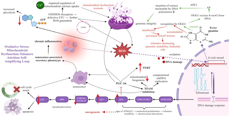
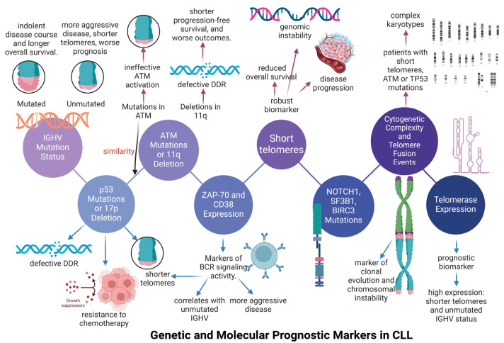
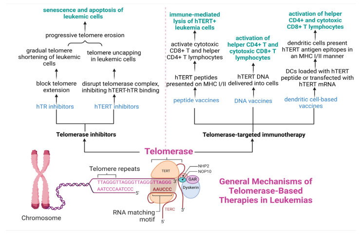
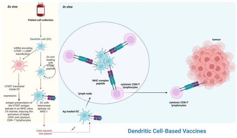
The nucleoprotein structures known as telomeres provide genomic integrity by protecting the ends of chromosomes. Tumorigenesis is associated with alterations in telomere function and stability. This narrative review provides evidence of the potential prognostic value of telomere length and telomerase in leukemias. On the one hand, oxidative stress and mitochondrial dysfunction can accelerate telomere shortening, leading to higher susceptibility and the progression of leukemia. On the other hand, cytogenetic alterations (such as gene fusions and chromosomal abnormalities) and genomic complexity can result from checkpoint dysregulation, the induction of the DNA damage response (DDR), and defective repair signaling at telomeres. This review thoroughly outlines the ways by which telomere dysfunction can play a key role in the development and progression of four primary leukemias, including chronic lymphocytic leukemia (CLL), chronic myeloid leukemia (CML), and acute leukemias of myeloid or lymphoid origin, highlighting the potential prognostic value of telomere length in this field. However, telomerase, which is highly active in leukemias, can prevent the rate of telomere attrition. In line with this, leukemia cells can proliferate, suggesting telomerase as a promising therapeutic target in leukemias. For this reason, telomerase-based immunotherapy is analyzed in the fight against leukemias, leveraging the immune system to eliminate leukemia cells with uncontrolled proliferation.

Keywords: genomic instability; immunotherapy in leukemia; leukemia; leukemia prognosis; mitochondrial dysfunction; oxidative stress; telomerase inhibitors; telomerase vaccines; telomerase-based therapy; telomere length.


Conflict of interest statement

The authors declare no conflicts of interest.

Figures

Figure 1
Figure 1
Oxidative stress–mitochondrial dysfunction–telomere attrition: A self-amplifying loop driving senescence and genomic instability. Reactive oxygen species (ROS) generated by defective oxidative phosphorylation (OXPHOS) impair the electron transport chain (ETC), further increasing ROS levels. ROS induces telomeric DNA oxidation, forming 8-oxo-guanine lesions, which disrupt the shelterin complex. This activates the DNA damage response (DDR) through ataxia telangiectasia mutated (ATM)/ataxia telangiectasia and Rad3-related (ATR) kinases, triggering checkpoint kinase 1 (CHK1)/checkpoint kinase 2 (CHK2), stabilizing p53, and upregulating p21, which inhibits cyclin-dependent kinase 4/6 (CDK4/6). This prevents retinoblastoma protein (Rb) phosphorylation, blocking E2F transcription factor activation and leading to cell cycle arrest, senescence, or apoptosis. Senescence-associated secretory phenotype (SASP) factors promote chronic inflammation, further amplifying oxidative stress. Mitochondrial dysfunction also suppresses peroxisome proliferator-activated receptor gamma coactivator 1-alpha/beta (PGC-1α/β), impairing mitochondrial biogenesis and energy metabolism. The loss of ATM or p53 function removes critical cell cycle checkpoints, leading to unchecked proliferation, chromosomal instability, and oncogenesis, reinforcing the loop (created with BioRender.com).
Figure 2
Figure 2
Genetic and molecular prognostic markers in Chronic Lymphocytic Leukemia (CLL). The immunoglobulin variable heavy-chain (IGHV) mutation status is a significant predictive factor, with mutated IGHV linked to indolent disease and unmutated IGHV associated with shorter telomeres and worse prognosis. Moreover, p53 mutations (17p deletion) and ataxia telangiectasia mutated (ATM) mutations (11q deletion) impair DNA damage response (DDR), leading to genomic instability, chemoresistance, and disease progression. Short telomeres could be biomarker of poor prognosis, driving chromosomal instability and clonal evolution. ZAP-70 and CD38 expression correlate with unmutated IGHV and aggressive disease, while NOTCH1, SF3B1, and BIRC3 mutations promote chromosomal instability and therapy resistance. Complex karyotypes and telomere fusion events are common in patients with ATM/p53 mutations, further worsening prognosis. Finally, high telomerase (hTERT) expression is linked to shorter telomeres, unmutated IGHV, and potential therapeutic targeting (created with BioRender.com).
Figure 3
Figure 3
The telomerase-based therapeutic options in leukemias. Telomerase inhibitors include hTR inhibitors that block telomere extension and hTERT inhibitors that disrupt telomerase complex formation, leading to telomere shortening or uncapping and triggering senescence or apoptosis of leukemic cells. In telomerase-targeted immunotherapy, peptide vaccines present hTERT antigens via major histocompatibility complex (MHC) I/II to activate CD8+ and CD4+ T cells. DNA vaccines deliver hTERT DNA to induce effector T cell responses. In dendritic cell-based vaccines, dendritic cells are loaded with hTERT peptides or transfected with hTERT mRNA, and they present hTERT epitopes to stimulate cytotoxic CD8+ and helper CD4+ T cells for killing leukemia cells (created with BioRender.com).
Figure 4
Figure 4
The molecular mechanisms underlying dendritic cell-based vaccines. This figure illustrates the mechanism of dendritic cell (DC)-based telomerase vaccines. Patient-derived DCs are either transfected with mRNA encoding human telomerase reverse transcriptase (hTERT) and lysosomal-associated membrane protein 1 (LAMP) or ex vivo loaded with hTERT peptide. Transfected DCs translate and present the telomerase epitope via major histocompatibility complex (MHC)-I and (MHC)-II, while peptide-loaded DCs present it via MHC-I. After injection into the patient, antigen-loaded DCs migrate to lymph nodes and activate cytotoxic CD8+ T lymphocytes, recognizing and killing hTERT-expressing tumor cells (created with BioRender.com).

Similar articles

References

    1. Hockemeyer D., Daniels J.-P., Takai H., de Lange T. Recent Expansion of the Telomeric Complex in Rodents: Two Distinct POT1 Proteins Protect Mouse Telomeres. Cell. 2006;126:63–77. doi: 10.1016/j.cell.2006.04.044. - DOI - PubMed
    1. Blackburn E.H., Epel E.S., Lin J. Human Telomere Biology: A Contributory and Interactive Factor in Aging, Disease Risks, and Protection. Science. 2015;350:1193–1198. doi: 10.1126/science.aab3389. - DOI - PubMed
    1. Palm W., De Lange T. How Shelterin Protects Mammalian Telomeres. Annu. Rev. Genet. 2008;42:301–334. doi: 10.1146/annurev.genet.41.110306.130350. - DOI - PubMed
    1. de Lange T. Shelterin: The Protein Complex That Shapes and Safeguards Human Telomeres. Genes Dev. 2005;19:2100–2110. doi: 10.1101/gad.1346005. - DOI - PubMed
    1. Apetroaei M.-M., Fragkiadaki P., Velescu B.Ș., Baliou S., Renieri E., Dinu-Pirvu C.E., Drăgănescu D., Vlăsceanu A.M., Nedea M.I.I., Udeanu D.I., et al. Pharmacotherapeutic Considerations on Telomere Biology: The Positive Effect of Pharmacologically Active Substances on Telomere Length. Int. J. Mol. Sci. 2024;25:7694. doi: 10.3390/ijms25147694. - DOI - PMC - PubMed

LinkOut - more resources