

Fluorescence Guidance in Glioma Surgery: A Narrative Review of Current Evidence and the Drive Towards Objective Margin Differentiation
- PMID: 40563668
- PMCID: PMC12190578
- DOI: 10.3390/cancers17122019
Fluorescence Guidance in Glioma Surgery: A Narrative Review of Current Evidence and the Drive Towards Objective Margin Differentiation
Abstract
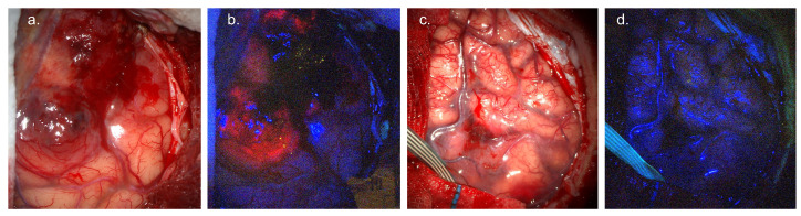
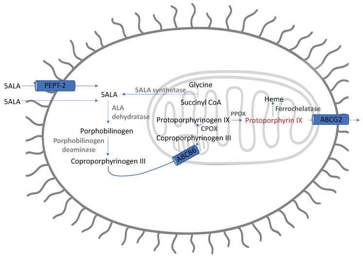
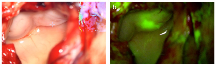
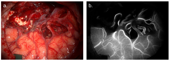
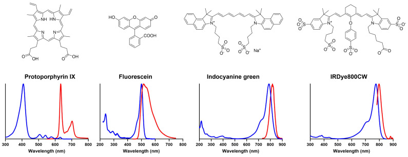
Fluorescence-guided surgery (FGS) was pioneered for glioma and is now established as the standard of care. Gliomas are infiltrative tumours with diffuse margins. FGS provides improved intra-operative identification of tumour margins based on tumour-specific emission visible to the operating surgeon, resulting in increased rates of gross total resection. Multiple fluorescence agents may be used including 5-ALA, fluorescein sodium, and indocyanine green (ICG). This review details the indication, required equipment, mechanism of action, evidence base, limitations, and regulatory issues for each fluorophore as utilised in current clinical practice. FGS for glioma is limited by a reliance on subjective interpretation of visible fluorescence, which is often not present in low-grade glioma (LGG) or at the infiltrative tumour margin. Consequently, there has been a drive to develop enhanced, objective FGS techniques utilising both quantitative fluorescence (QF) imaging systems and novel fluorophores. This review provides an overview of emerging QF imaging systems for FGS. The pipeline for novel fluorophore development is also summarised.
Keywords: 5-ALA; ICG; PpIX; fluorescein; fluorescence; fluorescence-guided surgery; glioma; quantitative fluorescence.
Conflict of interest statement
J.S. and T.V. are shareholders and co-founders of Hypervision Surgical Ltd., a King’s College London spinout company developing hyperspectral technology for surgical use. Hypervision surgical had no role in the review design, interpretation of data, or writing of this manuscript.
Figures
References
-
- Chaichana K.L., Jusue-Torres I., Navarro-Ramirez R., Raza S.M., Pascual-Gallego M., Ibrahim A., Hernandez-Hermann M., Gomez L., Ye X., Weingart J.D., et al. Establishing percent resection and residual volume thresholds affecting survival and recurrence for patients with newly diagnosed intracranial glioblastoma. Neuro-Oncol. 2014;16:113–122. doi: 10.1093/neuonc/not137. - DOI - PMC - PubMed
-
- Plaha P., Camp S., Cook J., McCulloch P., Voets N., Ma R., Taphoorn M.J., Dirven L., Grech-Sollars M., Watts C., et al. FUTURE-GB: Functional and ultrasound-guided resection of glioblastoma—A two-stage randomised control trial. BMJ Open. 2022;12:e064823. doi: 10.1136/bmjopen-2022-064823. - DOI - PMC - PubMed
Publication types
Grants and funding
LinkOut - more resources
Full Text Sources
