

Extracellular Vesicles and Purinergic Signaling in Alzheimer's Disease-Joining Forces for Novel Therapeutic Approach
- PMID: 40563742
- PMCID: PMC12190853
- DOI: 10.3390/brainsci15060570
Extracellular Vesicles and Purinergic Signaling in Alzheimer's Disease-Joining Forces for Novel Therapeutic Approach
Abstract
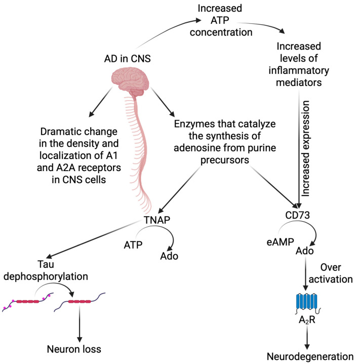
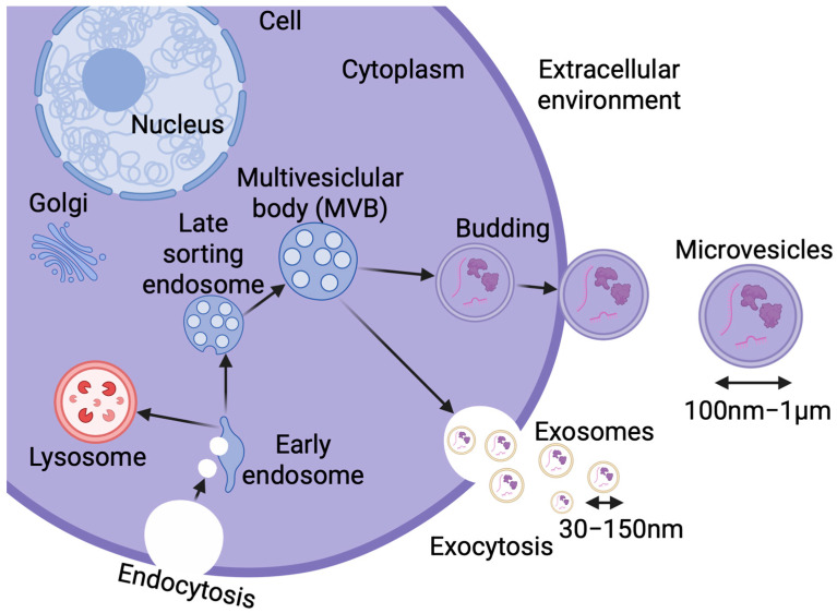
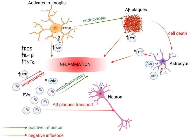
Neurodegenerative diseases, including Alzheimer's disease (AD), are a global problem affecting millions of people. Thanks to years of research and huge efforts, it has been possible to discover the pathophysiological changes accompanying Alzheimer's disease at the cellular level. It turns out that the formation of amyloid-beta plaques and hyperphosphorylation of tau protein in the brain play a key role in disease development. Purinergic signaling (PS) is implicated in the pathophysiology of several disorders in the central nervous system, and recent findings link some disturbances in PS with Alzheimer's disease. The primary objective of our review is to comprehensively explore and identify key purinergic signaling targets that hold therapeutic potential in the treatment of patients suffering from the disease. In particular, we focus on the dual role of purinergic compounds and extracellular vesicles (EVs), which have emerged as critical components in cellular communication and disease modulation. The extracellular vesicles that are naturally released by various cells fulfill the role of communication tools, also by harnessing the purinergic compounds. In this context, our review presents a thorough and integrative analysis of how extracellular vesicles can influence purinergic signaling and how this interaction might be leveraged to develop novel, targeted treatment strategies. Ultimately, this line of research may lead to innovative therapeutic approaches that are not only effective in slowing or halting disease progression but also demonstrate a high degree of biocompatibility and safety for the human organism.
Keywords: P2X receptors; extracellular vesicles; neurodegenerative diseases; neuroinflammation; purinergic signaling.
Conflict of interest statement
The authors declare no conflicts of interest.
Figures


Similar articles
-
CSF tau and the CSF tau/ABeta ratio for the diagnosis of Alzheimer's disease dementia and other dementias in people with mild cognitive impairment (MCI).Cochrane Database Syst Rev. 2017 Mar 22;3(3):CD010803. doi: 10.1002/14651858.CD010803.pub2. Cochrane Database Syst Rev. 2017. PMID: 28328043 Free PMC article.
-
Systemic pharmacological treatments for chronic plaque psoriasis: a network meta-analysis.Cochrane Database Syst Rev. 2021 Apr 19;4(4):CD011535. doi: 10.1002/14651858.CD011535.pub4. Cochrane Database Syst Rev. 2021. Update in: Cochrane Database Syst Rev. 2022 May 23;5:CD011535. doi: 10.1002/14651858.CD011535.pub5. PMID: 33871055 Free PMC article. Updated.
-
Factors that influence parents' and informal caregivers' views and practices regarding routine childhood vaccination: a qualitative evidence synthesis.Cochrane Database Syst Rev. 2021 Oct 27;10(10):CD013265. doi: 10.1002/14651858.CD013265.pub2. Cochrane Database Syst Rev. 2021. PMID: 34706066 Free PMC article.
-
How lived experiences of illness trajectories, burdens of treatment, and social inequalities shape service user and caregiver participation in health and social care: a theory-informed qualitative evidence synthesis.Health Soc Care Deliv Res. 2025 Jun;13(24):1-120. doi: 10.3310/HGTQ8159. Health Soc Care Deliv Res. 2025. PMID: 40548558
-
Systemic pharmacological treatments for chronic plaque psoriasis: a network meta-analysis.Cochrane Database Syst Rev. 2017 Dec 22;12(12):CD011535. doi: 10.1002/14651858.CD011535.pub2. Cochrane Database Syst Rev. 2017. Update in: Cochrane Database Syst Rev. 2020 Jan 9;1:CD011535. doi: 10.1002/14651858.CD011535.pub3. PMID: 29271481 Free PMC article. Updated.
References
-
- Albert M.S., DeKosky S.T., Dickson D., Dubois B., Feldman H.H., Fox N.C., Gamst A., Holtzman D.M., Jagust W.J., Petersen R.C., et al. The diagnosis of mild cognitive impairment due to Alzheimer’s disease: Recommendations from the National Institute on Aging-Alzheimer’s Association workgroups on diagnostic guidelines for Alzheimer’s disease. Alzheimers Dement. 2011;7:270–279. doi: 10.1016/j.jalz.2011.03.008. - DOI - PMC - PubMed
Publication types
LinkOut - more resources
Full Text Sources
