

Rapid Eye Movements in Sleep Furnish a Unique Probe into the Ontogenetic and Phylogenetic Development of the Visual Brain: Implications for Autism Research
- PMID: 40563746
- PMCID: PMC12190437
- DOI: 10.3390/brainsci15060574
Rapid Eye Movements in Sleep Furnish a Unique Probe into the Ontogenetic and Phylogenetic Development of the Visual Brain: Implications for Autism Research
Abstract
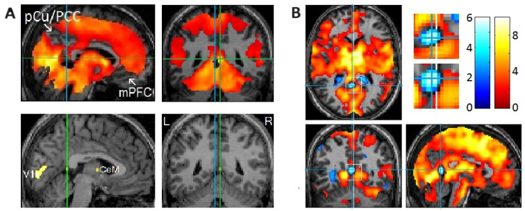
With positron emission tomography followed by functional magnetic resonance imaging (fMRI), we demonstrated that rapid eye movements (REMs) in sleep are saccades that scan dream imagery. The brain "sees" essentially the same way while awake and while dreaming in REM sleep. As expected, an event-related fMRI study (events = REMs) showed activation time-locked to REMs in sleep ("REM-locked" activation) in the oculomotor circuit that controls saccadic eye movements and visual attention. More crucially, the fMRI study provided a series of unexpected findings, including REM-locked multisensory integration. REMs in sleep index the processing of endogenous visual information and the hierarchical generation of dream imagery through multisensory integration. The neural processes concurrent with REMs overlap extensively with those reported to be atypical in autism spectrum disorder (ASD). Studies on ASD have shown atypical visual processing and multisensory integration, emerging early in infancy and subsequently developing into autistic symptoms. MRI studies of infants at high risk for ASD are typically conducted during natural sleep. Simply timing REMs may improve the accuracy of early detection and identify markers for stratification in heterogeneous ASD patients. REMs serve as a task-free probe useful for studying both infants and animals, who cannot comply with conventional visual activation tasks. Note that REM-probe studies would be easier to implement in early infancy because REM sleep, which is markedly preponderant in the last trimester of pregnancy, is still pronounced in early infancy. The brain may practice seeing the world during REM sleep in utero before birth. The REM-probe controls the level of attention across both the lifespan and typical-atypical neurodevelopment. Longitudinal REM-probe studies may elucidate how the brain develops the ability to "see" and how this goes awry in autism. REMs in sleep may allow a straightforward comparison of animal and human data. REM-probe studies of animal models of autism have great potential. This narrative review puts forth every reason to believe that employing REMs as a probe into the development of the visual brain will have far-reaching implications.
Keywords: animal model; autism spectrum disorders; functional MRI; multisensory integration; neurodevelopment; rapid eye movements in sleep; saccadic eye movements; visual perception.
Conflict of interest statement
The author declares no conflicts of interest.
Figures


Similar articles
-
Atypical development of sequential manual motor planning and visuomotor integration in children with autism at early school-age: A longitudinal kinematic study.Autism. 2025 Jun;29(6):1510-1523. doi: 10.1177/13623613241311333. Epub 2025 Jan 6. Autism. 2025. PMID: 39760319 Free PMC article.
-
Differently different?: A commentary on the emerging social cognitive neuroscience of female autism.Biol Sex Differ. 2024 Jun 13;15(1):49. doi: 10.1186/s13293-024-00621-3. Biol Sex Differ. 2024. PMID: 38872228 Free PMC article. Review.
-
REM Rebound Effect.2024 Sep 12. In: StatPearls [Internet]. Treasure Island (FL): StatPearls Publishing; 2025 Jan–. 2024 Sep 12. In: StatPearls [Internet]. Treasure Island (FL): StatPearls Publishing; 2025 Jan–. PMID: 32809548 Free Books & Documents.
-
Pharmacological intervention for irritability, aggression, and self-injury in autism spectrum disorder (ASD).Cochrane Database Syst Rev. 2023 Oct 9;10(10):CD011769. doi: 10.1002/14651858.CD011769.pub2. Cochrane Database Syst Rev. 2023. PMID: 37811711 Free PMC article.
-
Signs and symptoms to determine if a patient presenting in primary care or hospital outpatient settings has COVID-19.Cochrane Database Syst Rev. 2022 May 20;5(5):CD013665. doi: 10.1002/14651858.CD013665.pub3. Cochrane Database Syst Rev. 2022. PMID: 35593186 Free PMC article.
References
-
- Maenner M.J., Warren Z., Williams A.R., Amoakohene E., Bakian A.V., Bilder D.A., Durkin M.S., Fitzgerald R.T., Furnier S.M., Hughes M.M. Prevalence and characteristics of autism spectrum disorder among children aged 8 years—Autism and Developmental Disabilities Monitoring Network, 11 sites, United States, 2020. MMWR. Surveill. Summ. 2023;72:1–14. doi: 10.15585/mmwr.ss7202a1. - DOI - PMC - PubMed
-
- Ozonoff S., Young G.S., Carter A., Messinger D., Yirmiya N., Zwaigenbaum L., Bryson S., Carver L.J., Constantino J.N., Dobkins K. Recurrence risk for autism spectrum disorders: A Baby Siblings Research Consortium study. Pediatrics. 2011;128:e488–e495. doi: 10.1542/peds.2010-2825. - DOI - PMC - PubMed
Publication types
LinkOut - more resources
Full Text Sources
Research Materials
