. 2025 Jun 19;14(6):723.
doi: 10.3390/biology14060723.

Gut Microbiome Engineering for Diabetic Kidney Disease Prevention: A Lactobacillus rhamnosus GG Intervention Study

Affiliations

Gut Microbiome Engineering for Diabetic Kidney Disease Prevention: A Lactobacillus rhamnosus GG Intervention Study

Alaa Talal Qumsani. Biology (Basel). .

Abstract

The gut microbiota has emerged as a critical modulator in metabolic diseases, with substantial evidence supporting its role in attenuating diabetes-related nephropathy. Recent investigations demonstrate that strategic manipulation of intestinal microflora offers novel therapeutic avenues for safeguarding renal function against diabetic complications. This investigation sought to determine the nephroprotective potential of Lactobacillus rhamnosus GG (LGG) administration in diabetic nephropathy models. Six experimental cohorts were evaluated: control, probiotic-supplemented control, diabetic, diabetic receiving probiotic therapy, diabetic with antibiotics, and diabetic treated with both antibiotics and probiotics. Diabetic conditions were established via intraperitoneal administration of streptozotocin (50 mg/kg) following overnight fasting, according to validated protocols for experimental diabetes induction. Probiotic therapy (3 × 10⁹ CFU/kg, bi-daily) began one month before diabetes induction and continued throughout the study duration. Glycemic indices were monitored at bi-weekly intervals, inflammatory biomarkers, renal function indices, and urinary albumin excretion. The metabolic profile was evaluated through the determination of HOMA-IR and the computation of metabolic syndrome scores. Microbiome characterization employed 16S rRNA gene sequencing alongside metagenomic shotgun sequencing for comprehensive microbial community mapping. L. rhamnosus GG supplementation substantially augmented microbiome richness and evenness metrics. Principal component analysis revealed distinct clustering of microbial populations between treatment groups. The Prevotella/Bacteroides ratio, an emerging marker of metabolic dysfunction, normalized following probiotic intervention in diabetic subjects. Results: L. rhamnosus GG administration markedly attenuated diabetic progression, achieving glycated hemoglobin reduction of 32% compared to untreated controls. Pro-inflammatory cytokine levels (IL-6, TNF-α) decreased significantly, while anti-inflammatory mediators (IL-10, TGF-β) exhibited enhanced expression. The renal morphometric analysis demonstrated preservation of glomerular architecture and reduced interstitial fibrosis. Additionally, transmission electron microscopy confirmed the maintenance of podocyte foot process integrity in probiotic-treated groups. Conclusions: The administration of Lactobacillus rhamnosus GG demonstrated profound renoprotective efficacy through multifaceted mechanisms, including microbiome reconstitution, metabolic amelioration, and inflammation modulation. Therapeutic effects suggest the potential of a combined probiotic and pharmacological approach to attenuate diabetic-induced renal pathology with enhanced efficacy.

Keywords: Lactobacillus rhamnosus GG; diabetic nephropathy prevention; gut–kidney crosstalk; inflammatory regulation; metabolic modulation; microbiome engineering; reno protective mechanisms.


Conflict of interest statement

The authors declare that there are no conflicts of interest.

Figures

Figure 1
Figure 1
(A,B) Effects of Lactobacillus rhamnosus GG Supplementation on Body Weight and Glycemic Control in Diabetic Rats. (A) Weekly body weight measurements across experimental groups. (B) Fasting blood glucose levels over the study period. Subfigure (A) illustrates the longitudinal changes in body weight across experimental groups throughout the study period. While control animals maintained steady weight progression, diabetic rats exhibited marked weight reduction following alloxan-induced diabetes establishment. Administration of Lactobacillus rhamnosus GG alleviated but did not completely prevent the weight loss associated with diabetes. The prophylactic efficacy of probiotic supplementation was evidenced by significantly preserved body mass in treated diabetic animals compared to their untreated counterparts by study termination (** p < 0.01, *** p < 0.001). Subfigure (B) demonstrates the glycemic modulatory effects of L. rhamnosus GG, revealing markedly reduced fasting blood glucose (FBG) concentrations in probiotic-treated rats relative to diabetic controls (* p < 0.05). This substantial improvement in glycemic homeostasis underscores the metabolic benefits of targeted microbiome intervention.
Figure 2
Figure 2
(AF) Effects of Lactobacillus rhamnosus GG supplementation on glucose homeostasis and insulin dynamics in diabetic rats. This figure summarizes the impact of probiotic and/or metformin treatment on HOMA-IR, HOMA-β, AUC for OGTT and IST, and temporal glucose/insulin response curves at week 8. Data are presented as mean ± SD (n = 8–10/group). Statistical significance is indicated as follows: * p < 0.05, ** p < 0.01, vs. T2D control unless otherwise specified. Where applicable, inter-treatment comparisons are indicated within the figure.
Figure 3
Figure 3
(AG) Lactobacillus rhamnosus GG was used to study the effects of the bacteria on renal function and antioxidant biomarkers in diabetic rats. This figure depicts the influence that supplementation with L. rhamnosus GG, both on its own and in conjunction with metformin, had on the activities of antioxidant enzymes and kidney functional indicators in diabetic rats. Serum creatinine, blood urea nitrogen (BUN), urine albumin excretion, and the enzymatic activities of catalase (CAT), superoxide dismutase (SOD), and glutathione peroxidase (GPx) are among the parameters assessed. The statistics are provided as the mean plus the standard deviation, based on a sample size of eight to ten rats per group. The symbols * p < 0.05 and ** p < 0.01 are used to denote statistical significance in comparison to the diabetes group. Comparisons with the L. rhamnosus GG group are indicated by the symbols.
Figure 4
Figure 4
A comprehensive evaluation of renal functional parameters and histological changes following Lactobacillus rhamnosus GG intervention in diabetic nephropathy. Representative micrographs (AP) and quantitative analyses (QS) demonstrating the nephroprotective effects of L. rhamnosus GG in experimental diabetes. Control group (A) exhibits normal renal architecture with intact glomeruli and tubular structures. L. rhamnosus GG-supplemented control (B) shows preserved renal morphology similar to control. Diabetic kidneys (CF) display marked pathological alterations including glomerular hypertrophy, inflammatory cell infiltration, tubular damage, and interstitial fibrosis. Diabetic animals receiving metformin treatment (G,H) show moderate improvement in renal histopathology. Diabetic animals receiving L. rhamnosus GG treatment (I,J) display significant amelioration of histopathological features with preserved glomerular integrity and reduced inflammatory changes. Combined treatment with L. rhamnosus GG and metformin (K,L) demonstrates the most pronounced therapeutic effect with near-normal renal architecture. Higher magnification images (MP) reveal ultrastructural improvements in podocyte morphology and basement membrane thickness following single and combined interventions. Quantitative analyses confirm significant reductions in albuminuria (Q), glomerular filtration rate abnormalities (R), and renal tissue fibrosis (S) across treatment groups, with the most substantial improvements observed in the combined therapy group. The therapeutic hierarchy of efficacy is: T2D + L. rhamnosus GG + Metformin > T2D + L. rhamnosus GG > T2D + Metformin > T2D (untreated). Data are presented as the mean plus or minus the standard error of the mean (SEM), with a total of ten animals belonging to each group. The following equation was used to determine statistical significance: * p < 0.05, ** p < 0.01, in comparison to the control group; in comparison. In order to conduct the histological examination, panels (AP) were stained with hematoxylin and eosin (H&E). On each figure, the original magnification bar is shown.
Figure 4
Figure 4
A comprehensive evaluation of renal functional parameters and histological changes following Lactobacillus rhamnosus GG intervention in diabetic nephropathy. Representative micrographs (AP) and quantitative analyses (QS) demonstrating the nephroprotective effects of L. rhamnosus GG in experimental diabetes. Control group (A) exhibits normal renal architecture with intact glomeruli and tubular structures. L. rhamnosus GG-supplemented control (B) shows preserved renal morphology similar to control. Diabetic kidneys (CF) display marked pathological alterations including glomerular hypertrophy, inflammatory cell infiltration, tubular damage, and interstitial fibrosis. Diabetic animals receiving metformin treatment (G,H) show moderate improvement in renal histopathology. Diabetic animals receiving L. rhamnosus GG treatment (I,J) display significant amelioration of histopathological features with preserved glomerular integrity and reduced inflammatory changes. Combined treatment with L. rhamnosus GG and metformin (K,L) demonstrates the most pronounced therapeutic effect with near-normal renal architecture. Higher magnification images (MP) reveal ultrastructural improvements in podocyte morphology and basement membrane thickness following single and combined interventions. Quantitative analyses confirm significant reductions in albuminuria (Q), glomerular filtration rate abnormalities (R), and renal tissue fibrosis (S) across treatment groups, with the most substantial improvements observed in the combined therapy group. The therapeutic hierarchy of efficacy is: T2D + L. rhamnosus GG + Metformin > T2D + L. rhamnosus GG > T2D + Metformin > T2D (untreated). Data are presented as the mean plus or minus the standard error of the mean (SEM), with a total of ten animals belonging to each group. The following equation was used to determine statistical significance: * p < 0.05, ** p < 0.01, in comparison to the control group; in comparison. In order to conduct the histological examination, panels (AP) were stained with hematoxylin and eosin (H&E). On each figure, the original magnification bar is shown.
Figure 5
Figure 5
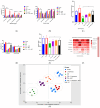
(A) Relative Abundance of Bacterial Phyla Across Experimental Groups. Stacked bar chart illustrating phylum-level taxonomic composition. Diabetic rats showed reduced Firmicutes (35%) and increased Bacteroidetes (35%) compared to controls (Firmicutes 75%). LGG supplementation partially restored Firmicutes abundance to 55%, indicating microbiome reconstitution. (B) Shannon Diversity Index of Gut Microbiota. Bar graph showing α-diversity across groups. Diabetic animals exhibited significantly reduced diversity (2.3 ± 0.28) versus controls (4.7 ± 0.12, p < 0.001). LGG-treated rats displayed significant improvement (4.0 ± 0.16), indicating recovery of microbial richness and evenness. (C) Firmicutes/Bacteroidetes Ratio as a Marker of Microbiome Balance. Bar graph depicting the F/B ratio, which was markedly reduced in diabetic rats (0.7 ± 0.18). LGG intervention significantly restored this ratio to 1.7 ± 0.11 (p < 0.01), indicating correction of gut dysbiosis. (D) Heatmap of Bacterial Phylum Abundance. Heatmap showing relative phylum-level abundances across groups. LGG administration resulted in Firmicutes recovery (55%) and normalization of minor phyla compared to diabetic rats (Firmicutes 35%), supporting microbiota-modulatory effects. (E) Principal Coordinate Analysis (PCoA) of Microbial Community Structure. PCoA plot based on weighted UniFrac distances shows distinct clustering of groups. LGG-treated microbiota clustered closer to controls, reflecting restored community architecture (explained variance: PC1 = 35%, PC2 = 27%). (F) Functional Potential of Gut Microbiota. Comparative bar chart indicating microbial functional profiles. LGG supplementation enhanced the expression of pathways involved in SCFA biosynthesis and carbohydrate metabolism, approaching control-like functional potential. (G) Compositional Analysis of Firmicutes and Bacteroidetes. Bar chart showing phylum-level proportions. Diabetic rats showed disrupted Firmicutes (35%) and Bacteroidetes (35%) balance. LGG restored Firmicutes to 55% and reduced Bacteroidetes to 25%, indicating microbiome modulation. Data are presented as mean ± SEM. Statistical significance was evaluated using one-way ANOVA followed by post hoc Tukey’s test. * p < 0.05, ** p < 0.01, *** p < 0.001 vs. T2D group.

Similar articles

References

    1. Gheith O., Farouk N., Nampoory N., Halim M., Al-Otaibi T. Diabetic kidney disease: Worldwide difference of prevalence and risk factors. J. Nephropharmacol. 2015;5:49. doi: 10.4103/1110-9165.197379. - DOI - PMC - PubMed
    1. Vaziri N.D., Wong J., Pahl M., Piceno Y.M., Yuan J., DeSantis T.Z., Ni Z., Nguyen T.H., Andersen G.L. Chronic kidney disease alters intestinal microbial flora. Kidney Int. 2013;83:308–315. doi: 10.1038/ki.2012.345. - DOI - PubMed
    1. Hiippala K., Jouhten H., Ronkainen A., Hartikainen A., Kainulainen V., Jalanka J., Satokari R. The potential of gut commensals in reinforcing intestinal barrier function and alleviating inflammation. Nutrients. 2018;10:988. doi: 10.3390/nu10080988. - DOI - PMC - PubMed
    1. Ishfaq M., Hu W., Hu Z., Guan Y., Zhang R. A review of nutritional implications of bioactive compounds of Ginger (Zingiber officinale Roscoe), their biological activities and nano-formulations. Ital. J. Food Sci. 2022;34:1–12. doi: 10.15586/ijfs.v34i3.2212. - DOI
    1. Fernández-Pampin N., González Plaza J.J., García-Gómez A., Peña E., Rumbo C., Barros R., Martel-Martín S., Aparicio S., Tamayo-Ramos J.A. Toxicology assessment of manganese oxide nanomaterials with enhanced electrochemical properties using human in vitro models representing different exposure routes. Sci. Rep. 2022;12:20991. doi: 10.1038/s41598-022-25483-w. - DOI - PMC - PubMed

LinkOut - more resources