Review
. 2025 May 23;13(6):1290.
doi: 10.3390/biomedicines13061290.

Ventricular Arrhythmias and Myocardial Infarction: Electrophysiological and Neuroimmune Mechanisms

Affiliations
Review

Ventricular Arrhythmias and Myocardial Infarction: Electrophysiological and Neuroimmune Mechanisms

Meng Zheng et al. Biomedicines. .

Abstract

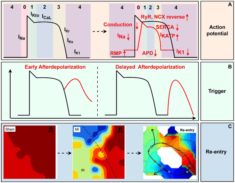
Ventricular arrhythmias (VAs) after myocardial infarction (MI) are still one of the most important causes of cardiovascular death, though patients receive timely vascular recanalization and drug treatment. And it requires further exploring the mechanism and new therapeutics of VAs induced by MI. Here, we review the electrophysiological and neuroimmune mechanisms of VAs induced by MI. Immune cells are regulated by combining with neuroendocrine molecules released by the sympathetic nervous system (SNS), and, in turn, they modulate SNS both at the paraventricular nucleus of the hypothalamus and stellate ganglion by releasing cytokines or chemokines. In addition, 'life essentials' such as sleep, physiological health, and exercise can also influence cardiovascular health through neuroimmune mechanisms. Those factors and mechanisms provide us with new perspectives for understanding the occurrence and maintenance of VAs after MI. Exploring the crosstalk between electrophysiology and neuroimmunology will contribute to finding new therapeutics for VAs after MI.

Keywords: crosstalk; immune system; myocardial infarction; sympathetic nervous system; ventricular arrhythmias.


Conflict of interest statement

The authors declare no conflicts of interest. The funders had no role in the design of the study; in the collection, analyses, or interpretation of data; in the writing of the manuscript; or in the decision to publish the results.

Figures

Figure 1
Figure 1
The electrophysiological mechanism of myocardial infarction. (A) RMP depolarized, and the amplitude and velocity of depolarization decreased, and APD shortened post-MI. (B) Early afterdepolarization occurs at the repolarization phase, while delayed afterdepolarization occurs after repolarization. (C) At the tissue level, reentrant ventricular arrhythmia originating from spatial heterogeneity and conduction block was recorded through optical mapping [20,21]. In the Sham and MI groups, red represents a short activation time, while blue represents a long activation time. In re - entry, red represents a long activation time, and blue represents a short activation time. RMP, rest membrane potential; RyR, ryanodine receptors; NCX, Na+/Ca2+-exchanger; SERCA, sarcoplasmic reticulum Ca2+-ATPase; APD, action potential duration; MI, myocardial infarction. (By Figdraw, https://www.figdraw.com/static/index.html#/, 11 March 2025).
Figure 2
Figure 2
The changes in sympathetic function in myocardial infarction. The increased cAMP-PKA/cGMP-PKG ratio exacerbated cardiac sympathetic activity, leading to Ca2+ influx into neurons, promoting neurotransmitters to be released. Meanwhile, MI causes transient cholinergic transdifferentiation of cardiac sympathetic nerves via gp130. The former exacerbates ventricular electrophysiological instability and has a pro-arrhythmic effect, while the latter may stabilize ventricular electrophysiological stability and has an anti-arrhythmic effect. The role of neurotransmitters on ventricular electrophysiology is indicated by the red arrow, and the blue arrow indicates the role of transdifferentiation on ventricular electrophysiology. SG, stellate ganglion; MI, myocardial infarction; VAs, ventricular arrhythmias; AP, action potential; NE, norepinephrine; NPY, Neuropeptide Y; GAL, galanin; ATP, adenosine triphosphate; APD, action potential duration; ERP, effective refractory period; VF, ventricular fibrillation. (By Figdraw, https://www.figdraw.com/static/index.html#/, 11 March 2025).
Figure 3
Figure 3
The neuroimmune mechanisms in myocardial Infarction. The regulation among the heart, the immune system, and the sympathetic nervous system (SNS) forms a complex network of cross-linking. On the one hand, the heart is regulated by the SNS from top to bottom and inflammation from the bone marrow (BM) and spleen. On the other hand, sympathetic nerves innervate both of them. From top to bottom, central sympathetic activity is influenced by various stimulating factors, and these stimuli can trigger microglial activation in the PVN area, thereby regulating central sympathetic neuron remodeling. The peripheral sympathetic ganglia are regulated by oxidative stress and the activation of satellite glial cells and inflammatory factors, resulting in an increase in sympathetic efferent that is relatively independent of central regulation. At the cardiac level, the myocardial tissue is affected by the neurotransmitters released by the efferent sympathetic nerve endings and the heterogeneous sympathetic nerve innervation, which exacerbates ventricular remodeling after MI. The immune cells derived from the resident myocardium, BM, and spleen can directly participate in the regulation of MI, on the one hand, and at the same time, they can also be regulated by the sympathetic nerve, thereby becoming a link in the middle of the SNS regulating MI. The solid black arrow represents a sympathetic efferent pathway; the black dotted arrow presents a visceral afferent pathway; the solid red arrow is an indirect effect of inflammation; PVN, paraventricular nucleus; BM, bone marrow; SG, sympathetic ganglia. (By Figdraw, https://www.figdraw.com/static/index.html#/, 11 March 2025).

Similar articles

Cited by

References

    1. Roth G.A., Mensah G.A., Johnson C.O., Addolorato G., Ammirati E., Baddour L.M., Barengo N.C., Beaton A.Z., Benjamin E.J., Benziger C.P., et al. Global Burden of Cardiovascular Diseases and Risk Factors, 1990–2019: Update From the GBD 2019 Study. J. Am. Coll. Cardiol. 2020;76:2982–3021. doi: 10.1016/j.jacc.2020.11.010. Erratum in J. Am. Coll. Cardiol. 2021, 77, 1958–1959. - DOI - PMC - PubMed
    1. Laforgia P.L., Auguadro C., Bronzato S., Durante A. The Reduction of Mortality in Acute Myocardial Infarction: From Bed Rest to Future Directions. Int. J. Prev. Med. 2022;13:56. doi: 10.4103/ijpvm.IJPVM_122_20. - DOI - PMC - PubMed
    1. Garcia R., Marijon E., Karam N., Narayanan K., Anselme F., Cesari O., Champ-Rigot L., Manenti V., Martins R., Puymirat E., et al. Ventricular fibrillation in acute myocardial infarction: 20-year trends in the FAST-MI study. Eur. Heart J. 2022;43:4887–4896. doi: 10.1093/eurheartj/ehac579. - DOI - PubMed
    1. Vallabhajosyula S., Patlolla S.H., Verghese D., Ya’Qoub L., Kumar V., Subramaniam A.V., Cheungpasitporn W., Sundaragiri P.R., Noseworthy P.A., Mulpuru S.K., et al. Burden of Arrhythmias in Acute Myocardial Infarction Complicated by Cardiogenic Shock. Am. J. Cardiol. 2020;125:1774–1781. doi: 10.1016/j.amjcard.2020.03.015. - DOI - PMC - PubMed
    1. Kosmidou I., Embacher M., McAndrew T., Dizon J.M., Mehran R., Ben-Yehuda O., Mintz G.S., Stone G.W. Early Ventricular Tachycardia or Fibrillation in Patients with ST Elevation Myocardial Infarction Undergoing Primary Percutaneous Coronary Intervention and Impact on Mortality and Stent Thrombosis (from the Harmonizing Outcomes with Revascularization and Stents in Acute Myocardial Infarction Trial) Am. J. Cardiol. 2017;120:1755–1760. doi: 10.1016/j.amjcard.2017.07.080. - DOI - PubMed

LinkOut - more resources