

Mechanisms Underlying Hyperexcitability: Combining Mossy Fiber Sprouting and Mossy Cell Loss in Neural Network Model of the Dentate Gyrus
- PMID: 40564135
- PMCID: PMC12190842
- DOI: 10.3390/biomedicines13061416
Mechanisms Underlying Hyperexcitability: Combining Mossy Fiber Sprouting and Mossy Cell Loss in Neural Network Model of the Dentate Gyrus
Abstract
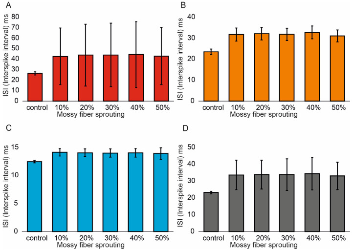
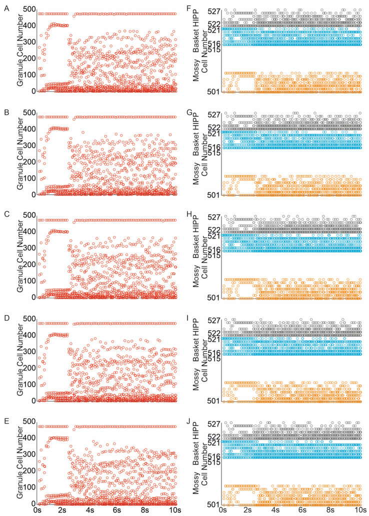
Background/Objectives: A concussive head injury increases the likelihood of temporal lobe epilepsy through mechanisms that are not entirely understood. This study aimed to investigate how two key histopathological features shared by both TLE (temporal lobe epilepsy) and head injury-mossy fiber sprouting and hilar excitatory cell loss-contribute to the modulation of dentate gyrus excitability. Methods: A computational approach was used to explore the impact of specific levels of mossy fiber sprouting and mossy cell loss, while avoiding the confounding effects of concurrent changes. The dentate gyrus model consists of 500 granule cells, 15 mossy cells, 6 basket cells and 6 hilar perforant path-associated cells. Results: My simulations demonstrate a correlation between the degree of mossy fiber sprouting and the number of spikes in dentate gyrus granule cells (correlations coefficient R = 0.95, p < 0.0001) and other cells (correlations coefficient R = 0.99, p < 0.0001). The mean values (standard deviation, SD) and 95% CI for granule cell activity in the control group and percentage 10-50% of mossy fiber sprouting groups are 376.4 (16.7) (95% CI, 374.9-377.8) vs. 463.5 (24.3) (95% CI, 461.4-465.6) vs. 514.8 (32.5) (95% CI, 511.9-517.6) vs. 555.0 (40.4) (95% CI, 551.5-558.6) vs. 633.4 (51.8) (95% CI, 628.8-637.9) vs. 701.7 (66.2) (95% CI, 695.9-707.5). The increase in mossy fiber sprouting was significantly statistically associated with an increase in granule cell activity (p < 0.01). The removal of mossy cells led to a reduction in excitability within the model network (for granule cells, correlations coefficient R = -0.40, p < 0.0001). Conclusions: These results are generally consistent with experimental observations, which indicate a high degree of mossy fiber sprouting in animals with a higher frequency of seizures. Whereas unlike the strong hyperexcitability effects induced by mossy fiber sprouting, the removal of mossy cells led to reduced granule cell responses to perforant path activation.
Keywords: dentate gyrus; epilepsy; mossy cell loss; mossy fiber sprouting; networks model.
Conflict of interest statement
The author declares no conflict of interest.
Figures


References
-
- Lowenstein D.H., Thomas M.J., Smith D.H., McIntosh T.K. Selective vulnerability of dentate hilar neurons following traumatic brain injury: A potential mechanistic link between head trauma and disorders of the hippocampus. J. Neurosci. 1992;12:4846–4853. doi: 10.1523/JNEUROSCI.12-12-04846.1992. - DOI - PMC - PubMed
LinkOut - more resources
Full Text Sources
