Review
. 2025 Jun 10;13(6):1426.
doi: 10.3390/biomedicines13061426.

Point-of-Care Ultrasound Use in Hemodynamic Assessment

Affiliations
Review

Point-of-Care Ultrasound Use in Hemodynamic Assessment

Ahmed Noor et al. Biomedicines. .

Abstract

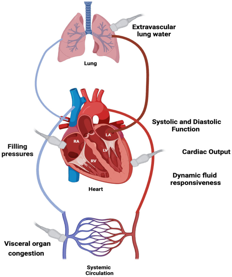
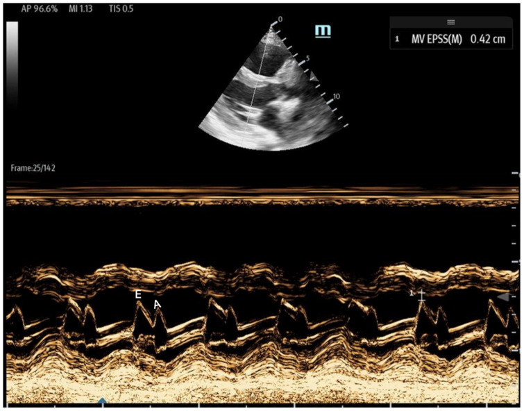
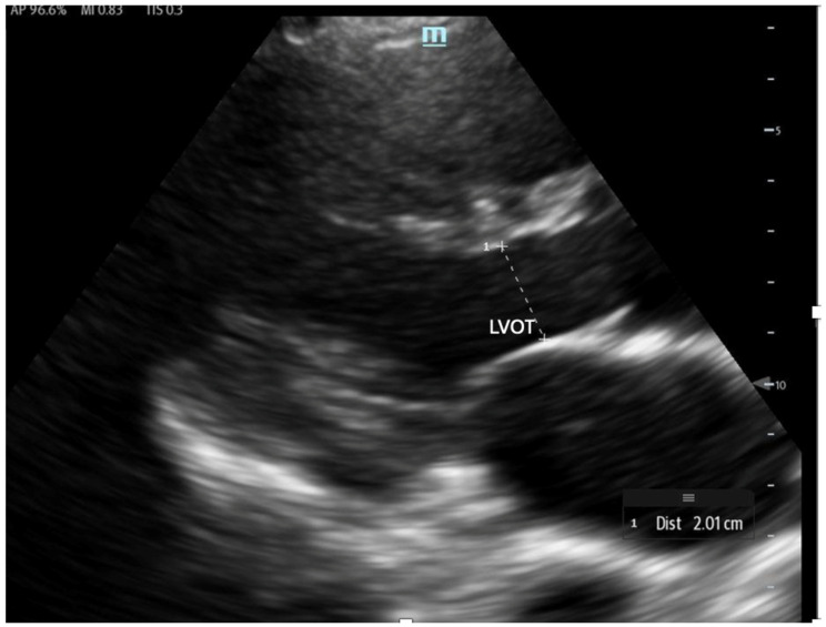
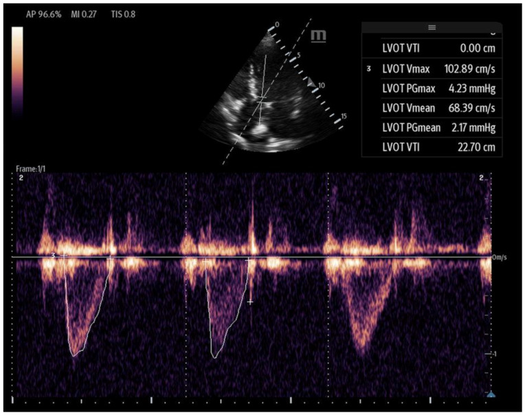
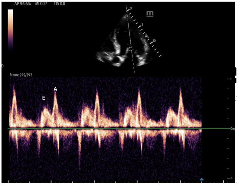
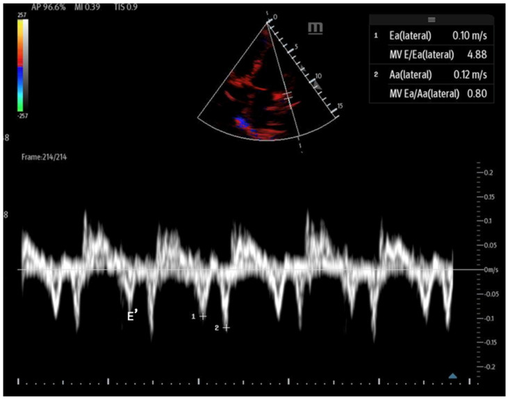
Hemodynamic assessment is critical in emergency and critical care for preventing, diagnosing, and managing shock states that significantly affect patient outcomes. Point-of-care ultrasound (POCUS) has become an invaluable, non-invasive, real-time, and reproducible tool for bedside decision-making. Advancements such as Doppler imaging, advanced critical care ultrasonography, and transesophageal echocardiography (TEE) have expanded its utility, enabling rapid and repeatable evaluations, especially in complex mixed shock presentations. This review explores the role of POCUS in hemodynamic monitoring, emphasizing its ability to assess cardiac output, filling pressures, and vascular congestion, facilitating shock classification and guiding fluid management. We highlight an extensive array of POCUS techniques for evaluating right and left cardiac function and review existing literature on their advantages, limitations, and appropriate clinical applications. Beyond assessing volume status, this review discusses the role of POCUS in predicting fluid responsiveness and supporting more individualized, precise management strategies. Ultimately, while POCUS is a powerful tool for rapid, comprehensive hemodynamic assessment in acute settings, its limitations must be acknowledged and thoughtfully integrated into clinical decision-making.

Keywords: cardiac arrest; critical care; echocardiography; emergency medicine; hemodynamics; point of care; ultrasound; volume assessment.


Conflict of interest statement

The authors declare no conflicts of interest.

Figures

Figure 1
Figure 1
Ultrasound integration in hemodynamic assessment evaluating cardiac output, fluid responsiveness, filling pressures, extravascular lung water, and visceral congestion.
Figure 2
Figure 2
The IVC (Inferior Vena Cava) diameter measured in subcoastal view approximately 1 cm below IVC–hepatic vein junction in a spontaneously breathing person during both inspiration (a) and expiration (b).
Figure 2
Figure 2
The IVC (Inferior Vena Cava) diameter measured in subcoastal view approximately 1 cm below IVC–hepatic vein junction in a spontaneously breathing person during both inspiration (a) and expiration (b).
Figure 3
Figure 3
Venous congestion evaluation using the ultrasound (VExUS) protocol for ultrasound scanning [24].
Figure 4
Figure 4
Diagrammatic representation of VExUS patterns: normal and abnormal patterns and grading for congestion.
Figure 5
Figure 5
Anechoic pericardial effusion in apical 4-chamber view.
Figure 6
Figure 6
Tricuspid annular plane systolic excursion (TAPSE): apical 4-chamber view with M-mode activation, demonstrating reduced TAPSE (measured at the bottom 2D M-mode).
Figure 7
Figure 7
A right ventricular tract VTI in a modified parasternal short axis view, activating the PW doppler.
Figure 8
Figure 8
(a) Tricuspid regurgitation velocity (TRV) measured by continuous wave (CW) Doppler in apical 4-chamber view. (b) D sign in parasternal short axis view in diastolic frame. Note flattening of the interventricular septum.
Figure 8
Figure 8
(a) Tricuspid regurgitation velocity (TRV) measured by continuous wave (CW) Doppler in apical 4-chamber view. (b) D sign in parasternal short axis view in diastolic frame. Note flattening of the interventricular septum.
Figure 9
Figure 9
Pulmonary acceleration time (PAT) measured in the subcoastal short axis, and PW doppler activation measured as the interval between the onset of pulmonary flow and peak velocity.
Figure 10
Figure 10
Example of B-lines, which are discrete vertical hyperechoic artifacts that originate at the pleural line and extend to the bottom of the screen.
Figure 11
Figure 11
The parasternal long-axis view with the M-mode shows the EPSS measurement, which is the minimal distance between the E wave (initial and maximal opening of the mitral for the passive filling of LV) and the septum. The E wave is followed by the A wave, which is smaller and corresponds to left atrial contraction.
Figure 12
Figure 12
The LVOT diameter measured below the opening of the aortic valve in a zoomed-in parasternal long axis to be incorporated into the LVOT area formula.
Figure 13
Figure 13
Left ventricular VTI calculated with PW Doppler with Doppler gate at LVOT.
Figure 14
Figure 14
Mitral inflow with PW Doppler gate at mitral valve tip (E and A waves).
Figure 15
Figure 15
Tissue Doppler at the lateral septal annulus to record the E′ wave (labeled).

Similar articles

References

    1. Kislitsina O.N., Rich J.D., Wilcox J.E., Pham D.T., Churyla A., Vorovich E.B., Ghafourian K., Yancy C.W. Shock—Classification and Pathophysiological Principles of Therapeutics. Curr. Cardiol. Rev. 2019;15:102–113. doi: 10.2174/1573403X15666181212125024. - DOI - PMC - PubMed
    1. Bakker J., Ince C. Monitoring coherence between the macro and microcirculation in septic shock. Curr. Opin. Crit. Care. 2020;26:267–272. doi: 10.1097/MCC.0000000000000729. - DOI - PubMed
    1. Caro C., Parker K. Mechanics and Imaging of the Macrocirculation. Magn. Reson. Med. 1990;14:179–186. doi: 10.1002/mrm.1910140204. - DOI - PubMed
    1. Khanna A., Karamchandani K. Macrocirculation and Microcirculation: The “Batman and Superman” Story of Critical Care Resuscitation. Anesth. Analg. 2021;132:280–283. doi: 10.1213/ANE.0000000000005272. - DOI - PubMed
    1. Ince C. Hemodynamic coherence and the rationale for monitoring the microcirculation. Crit. Care. 2015;19((Suppl. S3)):S8. doi: 10.1186/cc14726. - DOI - PMC - PubMed

LinkOut - more resources