. 2025 Jun 12;13(6):1447.
doi: 10.3390/biomedicines13061447.

Myricetin Amplifies Glucose-Stimulated Insulin Secretion via the cAMP-PKA-Epac-2 Signaling Cascade

Affiliations

Myricetin Amplifies Glucose-Stimulated Insulin Secretion via the cAMP-PKA-Epac-2 Signaling Cascade

Akhtar Ali et al. Biomedicines. .

Abstract

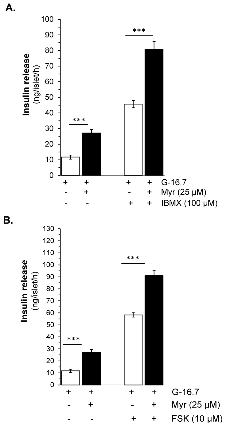
Aim: Myricetin, a natural bioflavonoid, is reported as an anti-diabetic agent since it possesses the ability to inhibit α-glucosidase activity, stimulate insulin action and secretion, manage ROS, and prevent diabetes complications. Myricetin was identified as a new insulin secretagogue that enhances glucose-stimulated insulin secretion and seems like a better antidiabetic drug candidate. Here, we explored the insulinotropic mechanism(s) of myricetin in vitro in mice islets and in silico. Methods: Size-matched pancreatic islets were divided into groups and incubated in the presence or absence of myricetin and agonists/antagonists of major insulin signaling pathways. The secreted insulin was measured by ELISA. Molecular docking studies were performed with the key player of insulin secretory pathways. Results: Myricetin dose-dependently enhanced insulin secretion in isolated mice islets, and its insulinotropic effect was exerted at high glucose concentrations distinctly different from glibenclamide. Myricetin-induced insulin secretion was significantly inhibited using the diazoxide. Furthermore, myricetin amplified glucose-induced insulin secretion in depolarized and glibenclamide-treated islets. Myricetin showed an additive effect with forskolin- and IBMX-induced insulin secretion. Interestingly, H89, a PKA inhibitor, and MAY0132, an Epac-2 inhibitor, significantly inhibited myricetin-induced insulin secretion. The in silico molecular docking studies further validated these in vitro findings in isolated pancreatic islets. Conclusions: Myricetin, a potential natural insulin secretagogue, amplifies glucose-induced insulin secretion via the cAMP-PKA-Epac-2 signaling pathway.

Keywords: antidiabetic drugs; insulin secretagogues; insulin secretion; myricetin; sulfonylurea.


Conflict of interest statement

The authors declare no conflicts of interest.

Figures

Figure 1
Figure 1
(A) Myricetin structure. (B) Myricetin enhances GSIS in isolated mice islets in a dose-dependent manner. Myricetin was added at 0, 1, 5, 25, and 100 μM concentrations in the presence of 3 mM or 16.7 mmol/L glucose. Myricetin does not induce insulin secretion at a 3 mM glucose concentration; however, at 16.7 mmol/L, a significant dose-dependent increase in insulin secretion was observed. * p < 0.05, ** p < 0.01, and *** p < 0.001 denote significant changes over the respective control values. The mean and SEM for the analysis were calculated based on five independent experiments conducted in triplicates.
Figure 2
Figure 2
Myricetin enhances glucose-dependent insulinotropic effect independent of K-ATP channels. (A) The effect of myricetin on insulin secretion in mice islets with K-ATP channels opened by diazoxide. Mice islets were incubated in 3 mM or 16.7 mM glucose in the presence or absence of myricetin and/or diazoxide (K-ATP channels opener), used at the indicated concentration. (B) Effect of myricetin on insulin secretion with Ca2⁺ channels blocked by verapamil. Islets were incubated in 3 mM or 16.7 mM glucose in the presence or absence of myricetin and/or verapamil at the indicated concentrations. The mean and SEM for the analysis were calculated based on five independent experiments conducted in triplicates. *** p < 0.001, significant changes over the respective control values.
Figure 3
Figure 3
Effects of myricetin on insulin secretion in (A), depolarized with Diazoxide- and KCl, and (B) Glibenclamide (GB)-treated mice islets. Islets were incubated in 16.7 mM glucose in the presence or absence of myricetin (25 μM) and/or diazoxide (50 μM) + KCl (25 mM) and GB (10 μM). The mean and SEM for the analysis were calculated based on 3 independent experiments conducted in triplicates. *** p < 0.001, significant changes over the respective control values.
Figure 4
Figure 4
Effects of myricetin on insulin secretion while inhibiting adenylyl cyclase. Islets were incubated in 16.7 mM glucose in the presence or absence of myricetin and/or SQ22536, an adenylyl cyclase inhibitor. The mean and SEM for the analysis were calculated based on 3 independent experiments conducted in triplicates. ** p < 0.01, significant changes when compared with myricetin alone.
Figure 5
Figure 5
Effect of myricetin on cAMP production and/or inhibition of cAMP hydrolysis. Effect of myricetin on insulin secretion (A) in the presence or absence of IBMX, a phosphodiesterase inhibitor, and/or (B) FSK, an adenylate cyclase activator. Islets were incubated in 16.7 mM glucose in the presence or absence of myricetin and/or IBMX/FSK at the indicated concentrations. The mean and SEM for the analysis were calculated based on 3 independent experiments conducted in triplicates. *** p < 0.001, significant changes when compared with control values.
Figure 6
Figure 6
PKA- and Epac2-dependent insulin secretory effect by myricetin. Islets were incubated at 16.7 mM glucose in the presence or absence of myricetin and (A) H-89, a PKA inhibitor, and (B) MAY 0132 (Epac-2 inhibitor). The mean and SEM for the analysis were calculated based on five independent experiments conducted in triplicates. *** p < 0.001, significant changes when compared with myricetin alone.
Figure 7
Figure 7
Molecular docking interactions of myricetin with various protein targets. Panel (A) illustrates the interaction of myricetin with Epac-2, (B) presents the key interactions of cognate ligand cAMP with Epac-2, (C) depicts the interaction of myricetin with the PKA RI alpha homodimer (PDB ID: 4MX3), and (D) shows the key residue interactions for cognate ligand cAMP of PKA RI alpha homodimer. Hydrogen bonds are indicated as blue solid lines, while hydrophobic interactions are represented as magenta dotted lines, emphasizing the binding interactions and conformational dynamics of myricetin with each respective protein target.
Figure 8
Figure 8
Effect of myricetin on the MEK kinase for the stimulation of insulin secretion. Islets were incubated in 16.7 mM glucose in the presence or absence of myricetin (A) U0126, a MEK kinase inhibitor, at the indicated concentrations. The mean and SEM for the analysis were calculated based on three independent experiments conducted in triplicates. *** p < 0.001 denotes significant changes when compared with Myr alone. Molecular docking interactions of myricetin with MEK-1 kinase. Panel (B) illustrates the interaction of myricetin with MEK kinase, (C) presents the 2D interactions of myricetin with MEK-1 kinase.
Figure 9
Figure 9
Model of myricetin-induced insulin secretory mechanisms. Myricetin amplifies GSIS in pancreatic islets through modulation of cAMP-PKA-Epac2 signaling cascade, independent of the direct involvement of KATP and Ca2+ channels. Continuous lines ending with arrows head indicate activation; continuous lines ending with block filled head indicate inhibition; dotted lines ending with arrows head indicate indirect involvement. cAMP, cyclic AMP; Epac, exchange protein activated by cAMP; PKA, protein kinase A; PKC, protein kinase C; PDE, phosphodiesterase; DAG, diacylglycerol; IP3, inositol 1,4,5-trisphosphate; RP, reserve pool; RRP, readily releasable pool; K+ -ATP channel, ATP-sensitive K+ channel.

Similar articles

References

    1. Xourafa G., Korbmacher M., Roden M.J. Inter-organ crosstalk during development and progression of type 2 diabetes mellitus. Nat. Rev. Endocrinol. 2024;20:27–49. doi: 10.1038/s41574-023-00898-1. - DOI - PubMed
    1. Bouchi R., Kondo T., Ohta Y., Goto A., Tanaka D., Satoh H., Yabe D., Nishimura R., Harada N., Kamiya H., et al. A consensus statement from the Japan Diabetes Society: A proposed algorithm for pharmacotherapy in people with type 2 diabetes. J. Diabetes Investig. 2023;14:151–164. doi: 10.1111/jdi.13960. - DOI - PMC - PubMed
    1. Mohajan D., Mohajan H.K. Oral Hypoglycaemic Agents: Non-Insulin Medications for Type 2 Diabetes Patients. Innov. Sci. Technol. 2024;3:23–31. doi: 10.56397/IST.2024.01.04. - DOI
    1. Kalwat M.A., Scheuner D., Rodrigues-dos-Santos K., Eizirik D.L., Cobb M.H. The pancreatic ß-cell response to secretory demands and adaption to stress. Endocrinology. 2021;162:bqab173. doi: 10.1210/endocr/bqab173. - DOI - PMC - PubMed
    1. Campbell J.E., Newgard C.B. Mechanisms controlling pancreatic islet cell function in insulin secretion. Nat. Rev. Mol. Cell Biol. 2021;22:142–158. doi: 10.1038/s41580-020-00317-7. - DOI - PMC - PubMed

LinkOut - more resources