

The Mechanism of Simvastatin-Mediated M1 Macrophage Polarization Contributing to Osteogenesis and Angiogenesis
- PMID: 40564174
- PMCID: PMC12190945
- DOI: 10.3390/biomedicines13061454
The Mechanism of Simvastatin-Mediated M1 Macrophage Polarization Contributing to Osteogenesis and Angiogenesis
Abstract
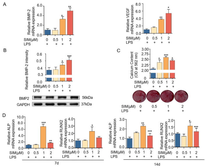
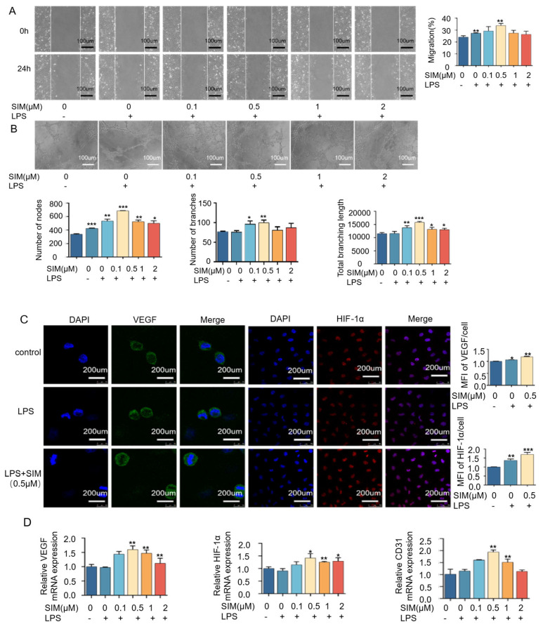
Background: The immune response is essential for bone regeneration, and macrophages in the immune microenvironment contribute to bone metabolism and angiogenesis. Emerging evidence demonstrates that simvastatin is a promising candidate for bone repair and promotes bone formation both in vitro and in vivo. However, the effect of simvastatin on macrophages and the following outcomes are still unclear. Objectives: This study aimed to investigate the potential immunomodulatory effect of simvastatin on M1 macrophages and its subsequent impact on osteogenesis and angiogenesis. Methods: Cell viability was assessed by CCK-8. Osteogenic and angiogenic markers were evaluated by RT-qPCR, Western blotting, and immunofluorescence. M1 macrophage phenotype was analyzed by flow cytometry. Osteogenesis was examined by histological staining, and angiogenic capacity was assessed using functional assays. Results: The present study found that simvastatin decreased M1 macrophage markers (CD86) and stimulated M1 macrophages to express high levels of pro-regenerative cytokines (BMP-2 and VEGF). In addition, simvastatin promoted osteogenic differentiation in MC3T3-E1 cells and angiogenic gene expression in HUVECs. Importantly, simvastatin enhanced the osteogenic capacity of MC3T3-E1 and the angiogenic potential of HUVECs by inhibiting M1 macrophage polarization in vitro. Conclusions: We demonstrated that simvastatin could confer favorable bone immunomodulatory properties and influence the crosstalk behavior between immune cells and osteoblasts and vascular endothelial cells to promote bone healing.
Keywords: angiogenesis; bone immune; macrophage polarization; osteogenesis; simvastatin.
Conflict of interest statement
The authors declare no conflicts of interest.
Figures


Similar articles
-
M2c Macrophages Mediate YAP1 to Promote Vascularized Bone Regeneration in Distraction Osteogenesis.FASEB J. 2025 Aug 15;39(15):e70923. doi: 10.1096/fj.202402895RR. FASEB J. 2025. PMID: 40788163 Free PMC article.
-
Receptor activator of nuclear factor-kappa B ligand-derived microglia healing peptide 1-AcN inhibits osteoarthritis progression in mice.Arthritis Res Ther. 2025 Jul 9;27(1):142. doi: 10.1186/s13075-025-03609-5. Arthritis Res Ther. 2025. PMID: 40635000 Free PMC article.
-
CX3CL1 promotes M1 macrophage polarization and osteoclast differentiation via NSUN5-mediated m5C modification.Sci Rep. 2025 Jul 12;15(1):25246. doi: 10.1038/s41598-025-11046-2. Sci Rep. 2025. PMID: 40652104 Free PMC article.
-
Anti-vascular endothelial growth factor for diabetic macular oedema: a network meta-analysis.Cochrane Database Syst Rev. 2017 Jun 22;6(6):CD007419. doi: 10.1002/14651858.CD007419.pub5. Cochrane Database Syst Rev. 2017. Update in: Cochrane Database Syst Rev. 2018 Oct 16;10:CD007419. doi: 10.1002/14651858.CD007419.pub6. PMID: 28639415 Free PMC article. Updated.
-
Role of Simvastatin on fracture healing and osteoporosis: a systematic review on in vivo investigations.Clin Exp Pharmacol Physiol. 2016 Jul;43(7):659-84. doi: 10.1111/1440-1681.12577. Clin Exp Pharmacol Physiol. 2016. PMID: 27061579
References
Grants and funding
LinkOut - more resources
Full Text Sources
