Review
. 2025 Jun 14;13(6):1471.
doi: 10.3390/biomedicines13061471.

Applications of Surface Plasmon Resonance in Heparan Sulfate Interactome Research

Affiliations
Review

Applications of Surface Plasmon Resonance in Heparan Sulfate Interactome Research

Payel Datta et al. Biomedicines. .

Abstract

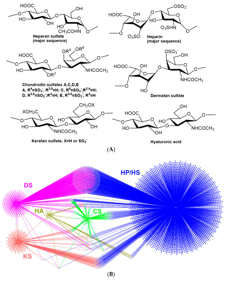
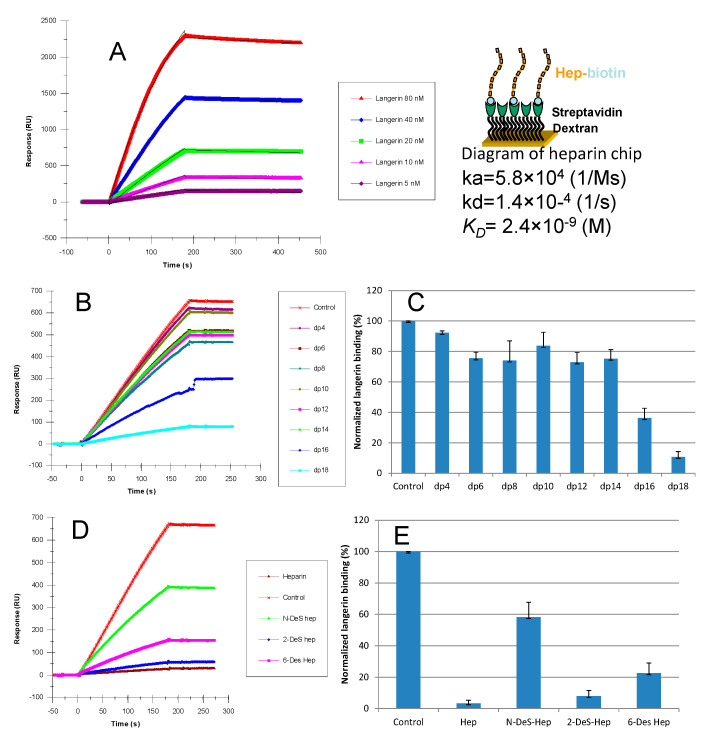
Surface plasmon resonance (SPR) is a powerful tool for analyzing biomolecular interactions and is widely used in basic biomedical research and drug discovery. Heparan sulfate (HS) is a linear complex polysaccharide and a key component of the extracellular matrix and cell surfaces. HS plays a pivotal role in maintaining cellular functions and tissue homeostasis by interacting with numerous proteins, making it essential for normal physiological processes and disease states. Deciphering the interactome of HS unlocks the mechanisms underlying its biological functions and the potential for novel HS-related therapeutics. This review presents an overview of the recent advances in the application of SPR technology to HS interactome research. We discuss methodological developments, emerging trends, and key findings that illustrate how SPR is expanding our knowledge of HS-mediated molecular interactions. Additionally, we highlight the potential of SPR-based approaches in identifying novel therapeutic targets and developing HS-mimetic drugs, thereby opening new avenues for intervention in HS-related diseases.

Keywords: heparan sulfate; heparin; interactome; surface plasmon resonance.


Conflict of interest statement

The authors declare no conflicts of interest.

Figures

Figure 1
Figure 1
(A) Disaccharide structures of major classes of GAGs. (B) The protein–GAGs interactome network, adapted from “The GAGs interactome 2.0” with permission. HP/HS, heparin/heparan sulfate (blue); CS, chondroitin sulfate (green); DS, dermatan sulfate (pink); HA, hyaluronic acid (dark yellow); KS, keratan sulfate (red). Each dot represents one protein binding partner. Some proteins (dots in light gray) can interact with multiple GAGs.
Figure 2
Figure 2
SPR has widespread applications in two major domains: (A) bioscience research, where SPR has been employed to investigate biomolecular interactions such as protein–protein, protein–DNA, polysaccharide–protein, and antibody–antigen binding; and (B) drug discovery and development, where SPR plays a crucial role in target identification and validation, high-throughput screening of potential drug candidates, lead optimization, and preclinical pharmacokinetic studies.
Figure 3
Figure 3
Typical reaction scheme for heparan sulfate and heparin biotinylation.
Figure 4
Figure 4
Typical SPR application for binding kinetics and structural analysis of heparin–protein interactions. (A) Left: SPR sensorgrams of langerin–heparin interaction; right: diagram of heparin chip and measured kinetics/affinity data for langerin–heparin interaction. Based on the sensorgrams, binding kinetics and affinity parameters (ka, kd, and KD) were calculated. (B) Sensorgrams of solution heparin oligosaccharides/surface heparin competition. (C) Bar graphs of normalized langerin binding preference to surface heparin by competing with different sizes of heparin oligosaccharides in solution, which shows the size dependence and minimum size of heparin oligosaccharide for the interaction. (D) Sensorgrams of solution chemical modified heparin/surface heparin competition. (E) Bar graphs of normalized langerin binding preference to surface heparin by competing with different chemically modified heparin in solution, which shows the sulfation dependence and sulfation preference of langerin–heparin interaction. Adapted from [52] with permission.
Figure 5
Figure 5
Solution competition SPR analysis of antithrombin (ATIII) and heparin interaction. (A) Diagram of SPR solution competition experiment for antithrombin (ATIII) binding to heparin. (B) SPR sensorgrams of ATIII binding to the heparin surface competing with different concentrations of heparin. The concentration of ATIII was 62.5 nM. Heparin concentrations in solution (from top to bottom) were 0, 3.13, 6.25, 12.5, 25, and 50 µg/mL, respectively. Adapted from [56] with permission.
Figure 6
Figure 6
IC50 measurement for the inhibition of sulfated glycans on the interactions between SARS-CoV-2 S-protein and heparin using SPR. (A) Competition SPR sensorgrams of SARS-CoV-2 S-protein and heparin interaction inhibited by different concentrations of heparin. (B) Dose–response curves for IC50 calculation of heparin using inhibition data from competition SPR. (C) Competition SPR sensorgrams of SARS-CoV-2 S-protein and heparin interaction inhibited by different concentrations of tri-sulfated HS. (D) Dose–response curves for IC50 calculation of tri-sulfated HS using inhibition data from competition SPR. Data based on our previous work [62] with permission.

Similar articles

References

    1. Nguyen H.H., Park J., Kang S., Kim M. Surface Plasmon Resonance: A Versatile Technique for Biosensor Applications. Sensors. 2015;15:10481. doi: 10.3390/s150510481. - DOI - PMC - PubMed
    1. Piliarik M., Vaisocherová H., Homola J. Surface Plasmon Resonance Biosensing. Methods Mol. Biol. 2009;503:65–88. doi: 10.1007/978-1-60327-567-5_5. - DOI - PubMed
    1. Zimmermann P., Egea-Jimenez A.L. Study of PDZ–Peptide and PDZ–Lipid Interactions by Surface Plasmon Resonance/BIAcore. Methods Mol. Biol. 2021;2256:75–87. doi: 10.1007/978-1-0716-1166-1_5. - DOI - PubMed
    1. Utt M., Danielsson B., Wadström T. Helicobacter Pylori Vacuolating Cytotoxin Binding to a Putative Cell Surface Receptor, Heparan Sulfate, Studied by Surface Plasmon Resonance. FEMS Immunol. Med. Microbiol. 2001;30:109–113. doi: 10.1111/j.1574-695X.2001.tb01557.x. - DOI - PubMed
    1. Zhang J., Liu B., Chen H., Zhang L., Jiang X. Application and Method of Surface Plasmon Resonance Technology in the Preparation and Characterization of Biomedical Nanoparticle Materials. Int. J. Nanomed. 2024;19:7049–7069. doi: 10.2147/IJN.S468695. - DOI - PMC - PubMed

LinkOut - more resources