. 2025 Jun 17;12(6):666.
doi: 10.3390/bioengineering12060666.

MRI-Based Machine Learning and Radiomics Methods for Assessing Spinal Cord Function in Patients with Mild Cervical Spondylotic Myelopathy

Affiliations

MRI-Based Machine Learning and Radiomics Methods for Assessing Spinal Cord Function in Patients with Mild Cervical Spondylotic Myelopathy

He Wang et al. Bioengineering (Basel). .

Abstract

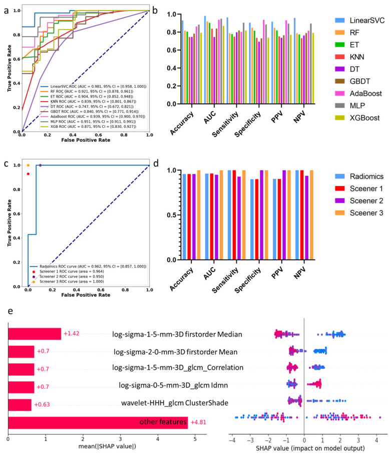
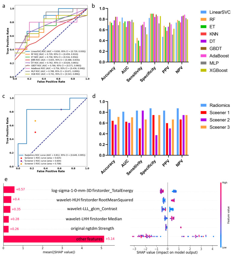
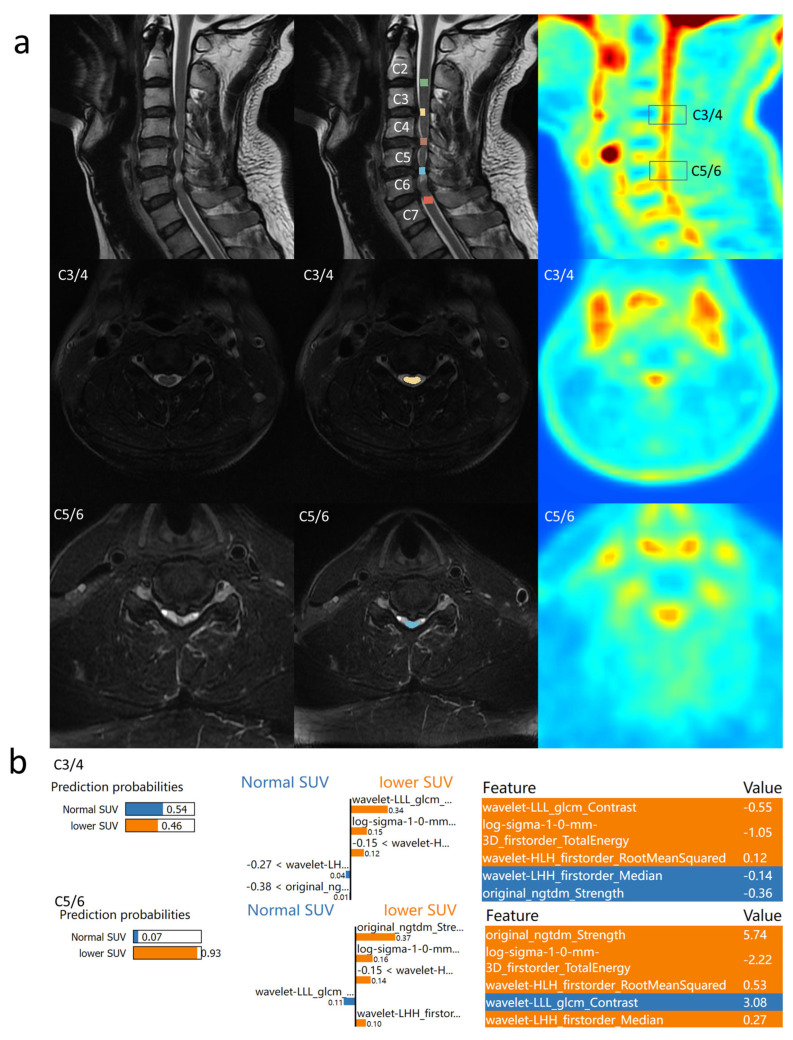
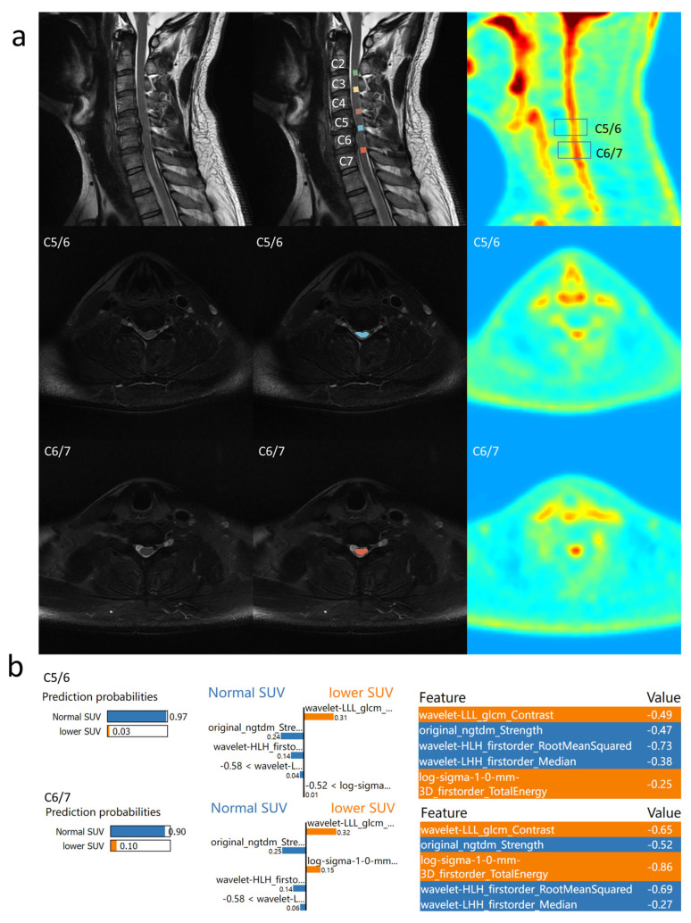
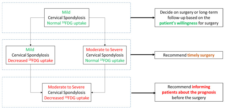
(1) Background: Patients with mild cervical spondylotic myelopathy (CSM) who delay surgery risk progression. While PET evaluates spinal cord function, its cost and radiation limit its use. (2) Methods: In this prospective study, patients with mild cervical spondylosis underwent preoperative 18F-FDG PET-MRI. Narrowed spinal levels were classified based on whether SUVmax was decreased. Follow-up assessments were conducted. Two machine learning models using MRI T2-based radiomics were developed to identify stenotic levels and decreased SUVmax. (3) Results: Patients with normal SUVmax showed greater symptom improvement. The radiomics models performed well, with AUCs of 0.981/0.962 (training/testing) for stenosis detection and 0.830/0.812 for predicting SUVmax decline. The model outperformed clinicians in predicting SUVmax decline, improving the AUC by 10%. (4) Conclusion: Patients with preserved SUVmax have better outcomes. MRI-based radiomics shows potential for identifying stenosis and predicting spinal cord function changes for preoperative assessment, though larger studies are needed to validate its clinical utility.

Keywords: PET-MRI; cervical spondylotic myelopathy; machine learning; radiomics.


Conflict of interest statement

The authors have no conflicts of interest to declare.

Figures

Figure 1
Figure 1
Results of identifying compression levels: (a) AUC curve of 9 machine learning algorithms on the training dataset; (b) statistic comparison of 9 machine learning algorithms on the training dataset; (c) AUC curve of proposed machine learning algorithm and 3 screeners on the test dataset; (d) statistic comparison of proposed machine learning algorithm and 3 screeners on the test dataset; (e) SHAPLEY explanations of the proposed model.
Figure 2
Figure 2
Results of identifying decreased 18F-FDG uptake levels: (a) AUC curve of 9 machine learning algorithms on the training dataset; (b) statistic comparison of 9 machine learning algorithms on the training dataset; (c) AUC curve of proposed machine learning algorithm and 3 screeners on the test dataset; (d) statistic comparison of proposed machine learning algorithm and 3 screeners on the test dataset; (e) SHAPLEY explanations of the proposed model.
Figure 3
Figure 3
Prediction result of Example 1: (a) the PET/MR results of Example 1 are shown. Cervical spinal cord segments from C2 to C7 and five intervertebral disc segments were labeled. SUVmax was normal at the C3/4 level and SUVmax was decreased at the C5/6 level; (b) the LIME results of the proposed model are shown.
Figure 4
Figure 4
Prediction result of Example 2: (a) the PET/MR results of Example 2 are shown. Cervical spinal cord segments from C2 to C7 and five intervertebral disc segments were labeled. SUVmax decreased at the C5/6 level and SUVmax was normal at the C6/7 level; (b) the LIME results of the proposed model are shown. The proposed methods predicted that both SUVmax were normal at the two levels.
Figure 5
Figure 5
Proposed flowchart of the treatment of cervical spondylotic myelopathy.

Similar articles

References

    1. Badhiwala J.H., Ahuja C.S., Akbar M.A., Witiw C.D., Nassiri F., Furlan J.C., Curt A., Wilson J.R., Fehlings M.G. Degenerative cervical myelopathy—Update and future directions. Nat. Rev. Neurol. 2020;16:108–124. doi: 10.1038/s41582-019-0303-0. - DOI - PubMed
    1. Fehlings M.G., Kwon B.K., Tetreault L.A. Guidelines for the management of degenerative cervical myelopathy and spinal cord injury: An introduction to a focus issue. Glob. Spine J. 2017;7:6S–7S. doi: 10.1177/2192568217701714. - DOI - PMC - PubMed
    1. Karadimas S.K., Erwin W.M., Ely C.G., Dettori J.R., Fehlings M.G. Pathophysiology and natural history of cervical spondylotic myelopathy. Spine. 2013;38:S21–S36. doi: 10.1097/BRS.0b013e3182a7f2c3. - DOI - PubMed
    1. Rhee J., Tetreault L.A., Chapman J.R., Wilson J.R., Smith J.S., Martin A.R., Dettori J.R., Fehlings M.G. Nonoperative versus operative management for the treatment degenerative cervical myelopathy: An updated systematic review. Glob. Spine J. 2017;7((Suppl. 3)):35S–41S. doi: 10.1177/2192568217703083. - DOI - PMC - PubMed
    1. Aiello M., Alfano V., Salvatore E., Cavaliere C., Picardi M., Della Pepa R., Nicolai E., Soricelli A., Vella A., Salvatore M., et al. [18F] FDG uptake of the normal spinal cord in PET/MR imaging: Comparison with PET/CT imaging. EJNMMI Res. 2020;10:91. doi: 10.1186/s13550-020-00680-8. - DOI - PMC - PubMed

LinkOut - more resources