

19-Gauge Versus 22-Gauge Franseen Needles, Comparison of the Histological Diagnostic Capability of Endoscopic Ultrasound-Guided Fine-Needle Biopsy for Autoimmune Pancreatitis: A Multicenter Retrospective Cohort Study
- PMID: 40564818
- PMCID: PMC12192491
- DOI: 10.3390/diagnostics15121496
19-Gauge Versus 22-Gauge Franseen Needles, Comparison of the Histological Diagnostic Capability of Endoscopic Ultrasound-Guided Fine-Needle Biopsy for Autoimmune Pancreatitis: A Multicenter Retrospective Cohort Study
Abstract
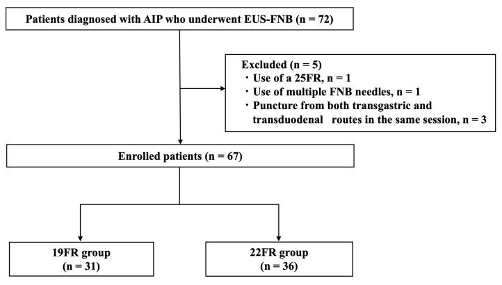
Background/Objectives: Endoscopic ultrasound-guided fine-needle biopsy (EUS-FNB) is a useful procedure for obtaining histological specimens. However, its utility in diagnosing autoimmune pancreatitis (AIP) has not yet been well studied. This study aimed to assess the diagnostic capability of EUS-FNB for AIP by comparing a 19-gauge Franseen needle (19FR) and a 22-gauge Franseen needle (22FR). Methods: This study included patients with a final diagnosis of AIP undergoing EUS-FNB for pancreatic lesions between January 2014 and February 2023. All patients underwent EUS-FNB with either 19FR or 22FR. Histological findings were evaluated according to the International Consensus Diagnostic Criteria (ICDC). The primary outcome was the diagnostic yield of Level 1 (≥3 ICDC items) or Level 2 (2 ICDC items). Results: The 19FR group included 31 patients, and the 22FR group included 36 patients. The Level 1 diagnostic rate was significantly higher in the 19FR group than in the 22FR group (90.3% vs. 61.1%, p = 0.010). No significant difference was observed in the Level 2 diagnostic rate. The 19FR group yielded significantly larger histological tissue samples than the 22FR group (median area: 9.19 mm2/session vs. 3.36 mm2/session, p < 0.001). The analysis demonstrated a positive correlation between tissue area and the number of histological diagnostic items obtained. Conclusions: EUS-FNB performed with the 19FR provided larger histological specimens and a higher histological diagnostic yield than the 22FR in the diagnosis of AIP. Obtaining a larger amount of tissue may facilitate a definitive diagnosis of AIP.
Keywords: 19-gauge; 22-gauge; Franseen needle; autoimmune pancreatitis; endoscopic ultrasound-guided fine needle biopsy.
Conflict of interest statement
The authors declare no conflicts of interest.
Figures


Similar articles
-
Endoscopic ultrasound-guided fine-needle biopsy needle can facilitate histological diagnosis of type 1 autoimmune pancreatitis.J Hepatobiliary Pancreat Sci. 2025 Mar;32(3):238-245. doi: 10.1002/jhbp.12095. Epub 2024 Dec 6. J Hepatobiliary Pancreat Sci. 2025. PMID: 39639754
-
Usefulness of endoscopic ultrasound-guided fine-needle biopsy for the diagnosis of autoimmune pancreatitis using a 22-gauge Franseen needle: a prospective multicenter study.Endoscopy. 2020 Nov;52(11):978-985. doi: 10.1055/a-1183-3583. Epub 2020 Jun 24. Endoscopy. 2020. PMID: 32583394 Clinical Trial.
-
EUS FNAC without rapid on-site evaluation is comparable to EUS FNB with macroscopic on-site evaluation in evaluation of intra-abdominal masses.Indian J Gastroenterol. 2025 Jun;44(3):371-377. doi: 10.1007/s12664-025-01741-3. Epub 2025 Feb 19. Indian J Gastroenterol. 2025. PMID: 39969684
-
Comparative diagnostic performance of end-cutting fine-needle biopsy needles for EUS tissue sampling of solid pancreatic masses: a network meta-analysis.Gastrointest Endosc. 2022 Jun;95(6):1067-1077.e15. doi: 10.1016/j.gie.2022.01.019. Epub 2022 Feb 4. Gastrointest Endosc. 2022. PMID: 35124072
-
Diagnostic yield of endoscopic and EUS-guided biopsy techniques in subepithelial lesions of the upper GI tract: a systematic review.Gastrointest Endosc. 2024 Jun;99(6):895-911.e13. doi: 10.1016/j.gie.2024.02.003. Epub 2024 Feb 13. Gastrointest Endosc. 2024. PMID: 38360118
Cited by
-
Endoscopic Diagnostics for IgG4-Related Pancreatobiliary Diseases: Current Modalities and Clinical Perspectives.Diagnostics (Basel). 2025 Aug 8;15(16):1990. doi: 10.3390/diagnostics15161990. Diagnostics (Basel). 2025. PMID: 40870842 Free PMC article. Review.
References
-
- Shimosegawa T., Chari S.T., Frulloni L., Kamisawa T., Kawa S., Mino-Kenudson M., Kim M.H., Klöppel G., Lerch M.M., Löhr M., et al. International Consensus Diagnostic Criteria for Autoimmune Pancreatitis Guidelines of the International Association of Pancreatology. Pancreas. 2011;40:352–358. doi: 10.1097/MPA.0b013e3182142fd2. - DOI - PubMed
-
- The Japanese Pancreas Society. the Research Committee of Intractable Disease for IgG4-Related Disease Supported by the Japanese Ministry of Health, Labor and Welfare Amendment of the Japanese Consensus Guidelines for Autoimmune Pancreatitis, 2020. Suizo. 2020;35:465–550. doi: 10.2958/suizo.35.465. (In Japanese) - DOI
LinkOut - more resources
Full Text Sources
Miscellaneous
