Review
. 2025 Jun 13;15(12):1508.
doi: 10.3390/diagnostics15121508.

The Hôtel-Dieu MRI Classification of Uterosacral Ligament Involvement in Endometriosis: A Pictorial Guide to Clinical Use

Affiliations
Review

The Hôtel-Dieu MRI Classification of Uterosacral Ligament Involvement in Endometriosis: A Pictorial Guide to Clinical Use

Siegfried Hélage et al. Diagnostics (Basel). .

Abstract

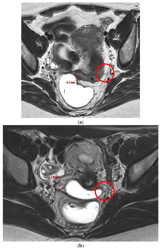
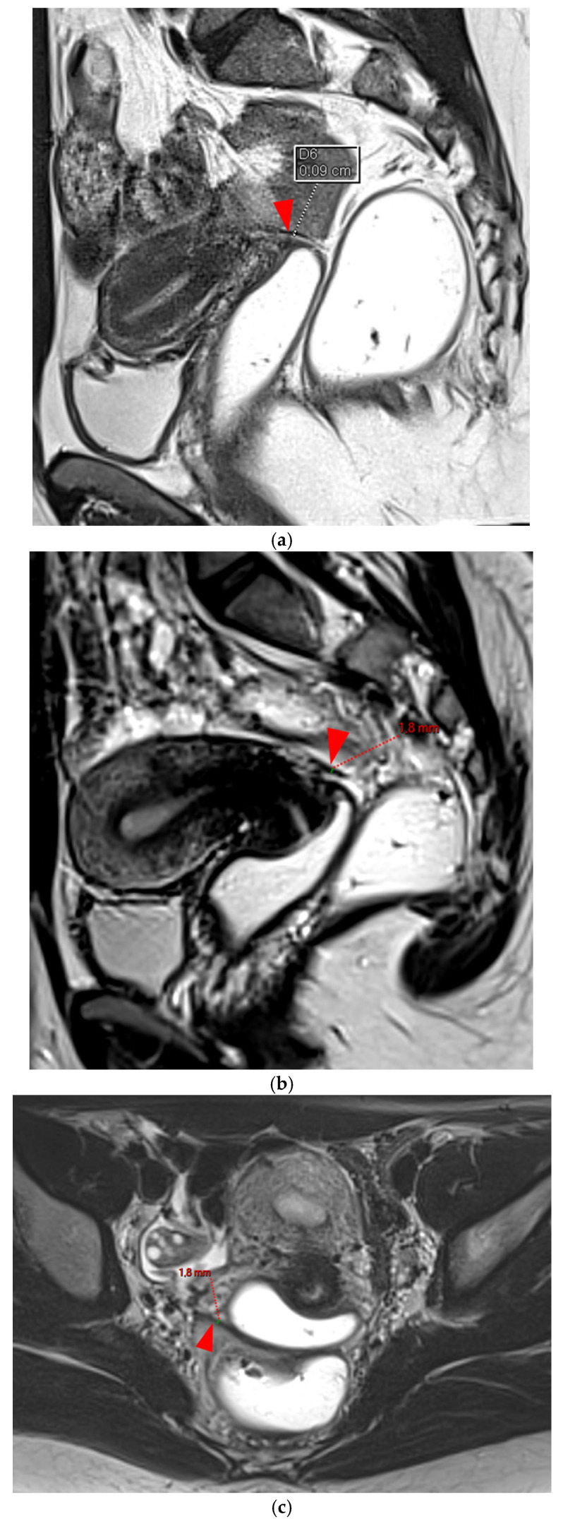
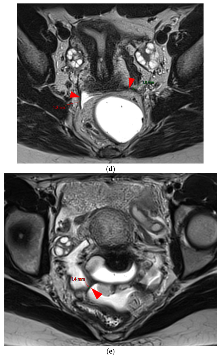
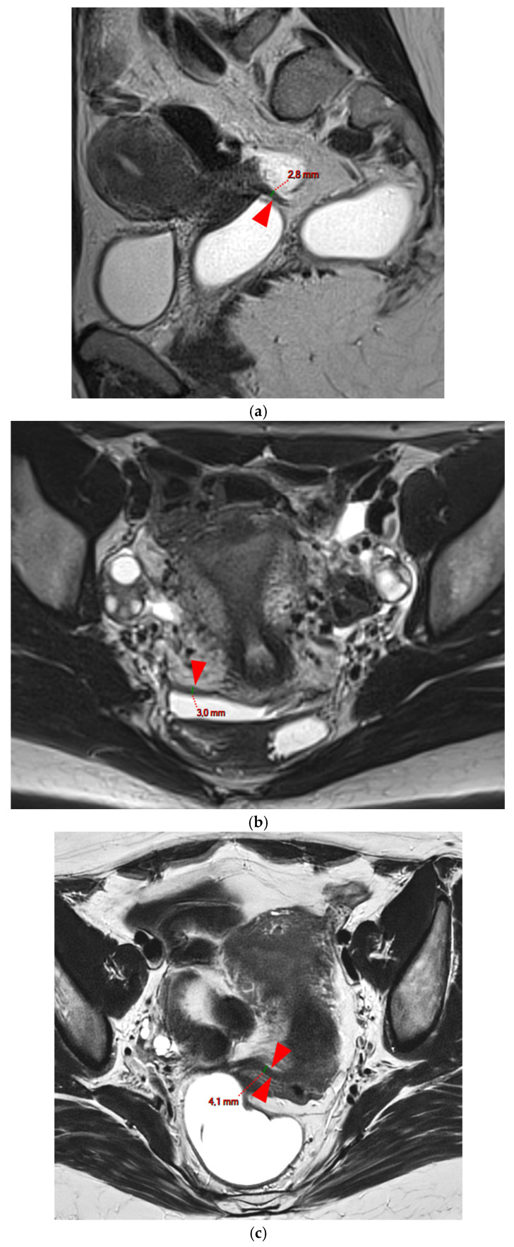
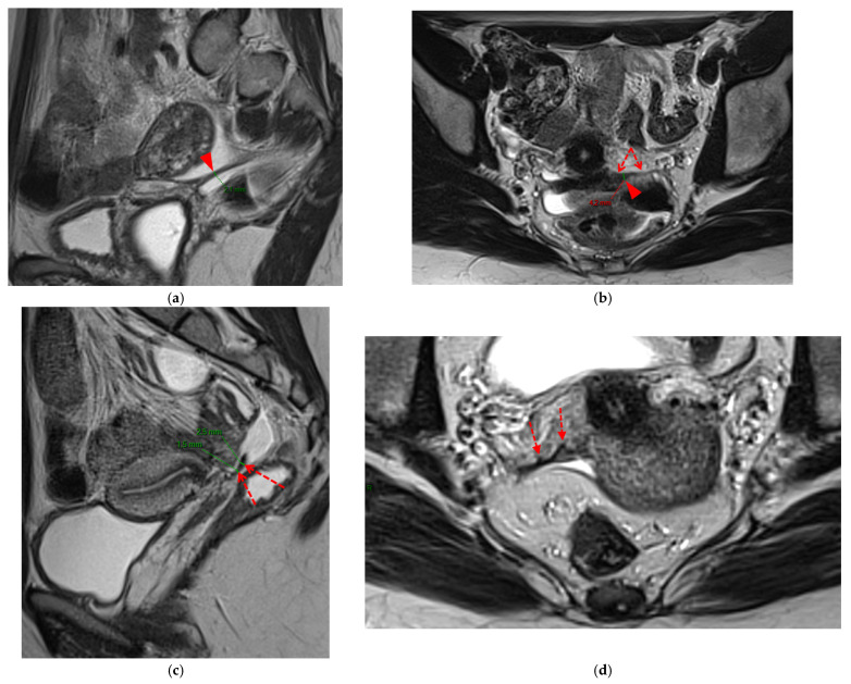
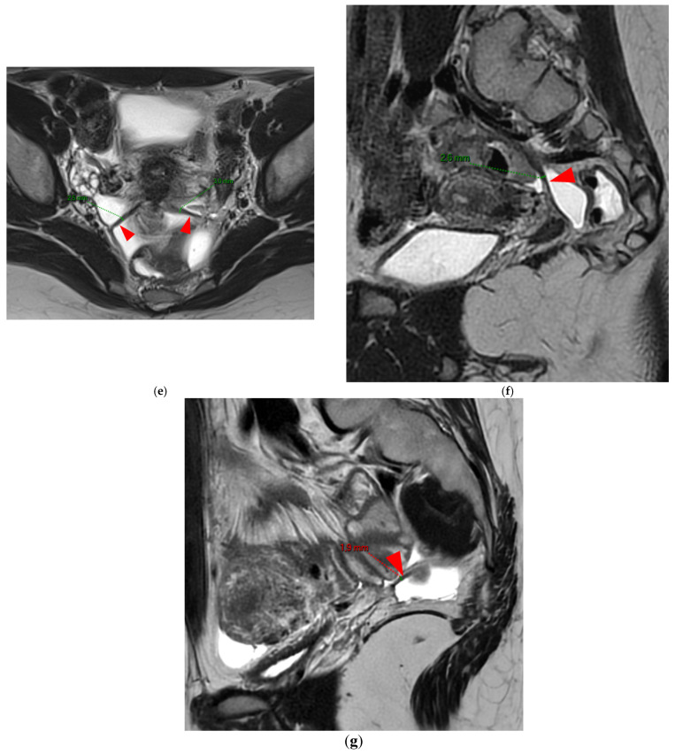
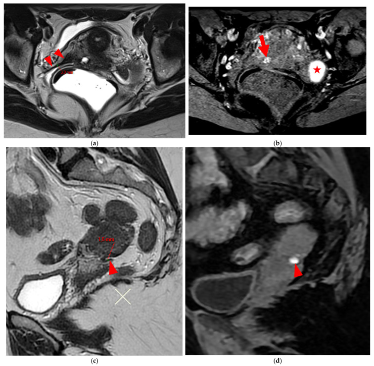
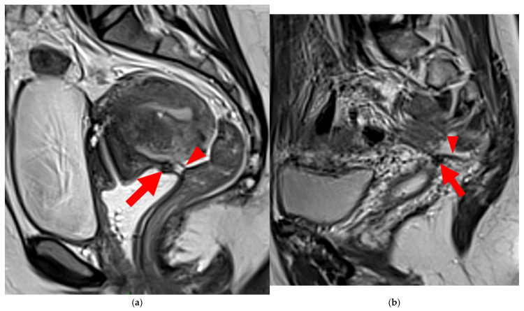
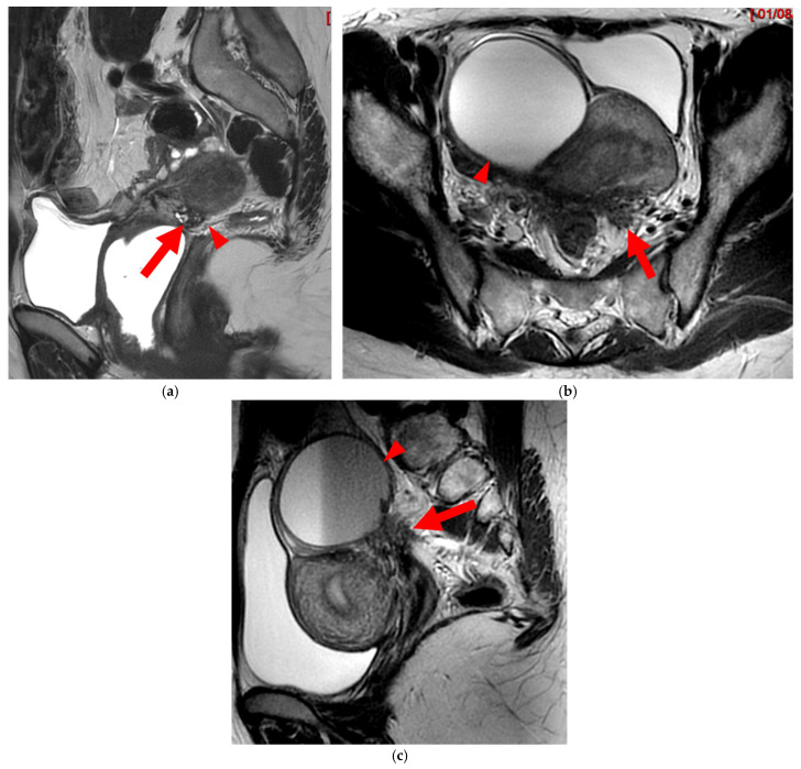
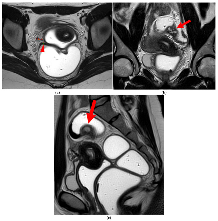
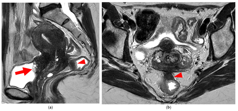
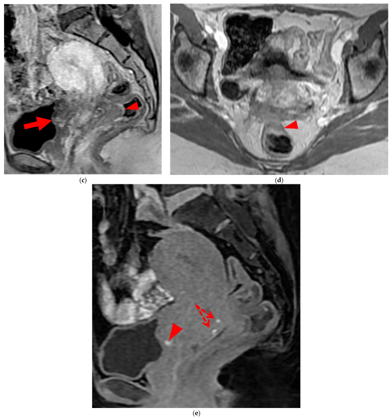
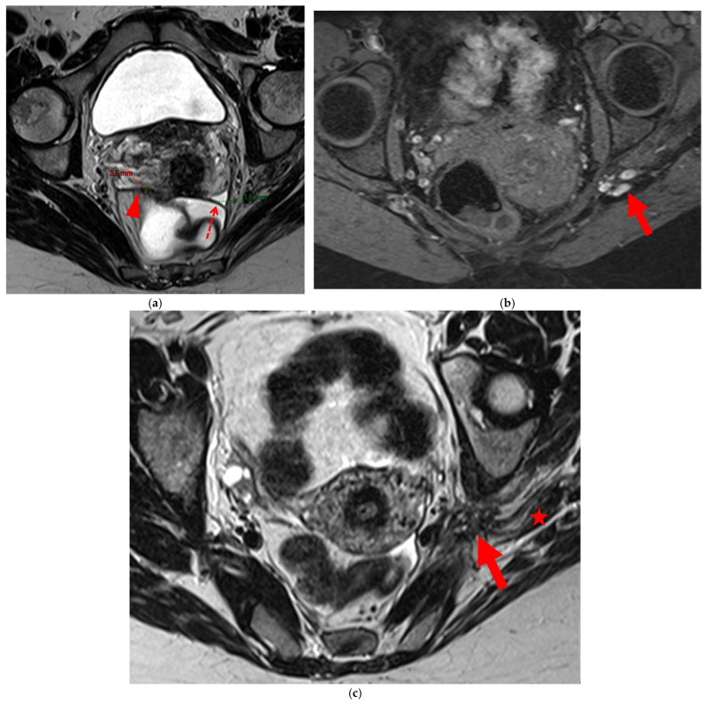
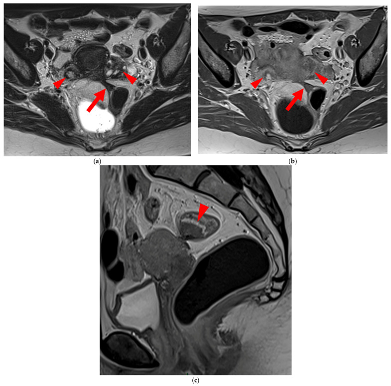
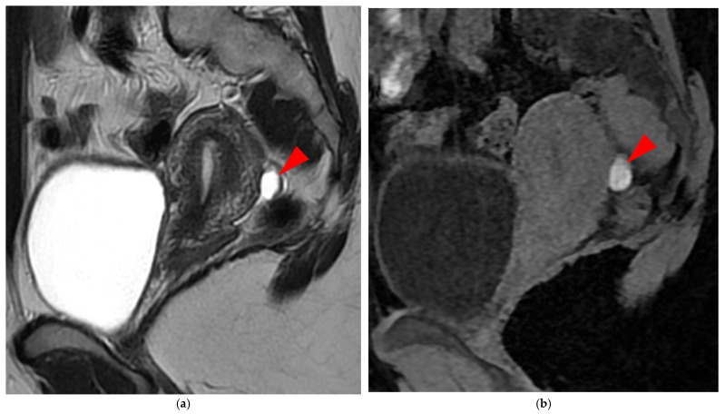
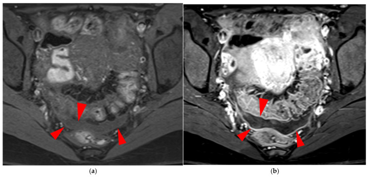
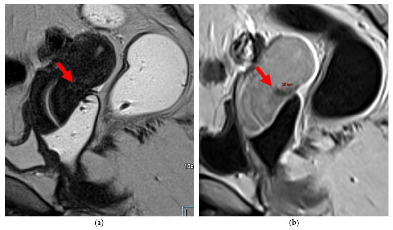
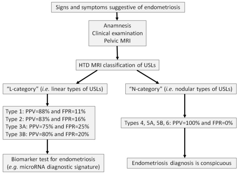
Objectives: Endometriosis is a common gynecologic condition characterized by the presence of endometrial-like tissue outside the uterus, often leading to pelvic pain and infertility. Diagnosis is frequently delayed, with prolonged diagnostic wandering that could be improved through enhanced first-line radiologic assessment. The uterosacral ligament (USL) is the most frequent site of deep infiltrating endometriosis (DIE). The Hôtel-Dieu (HTD) MRI classification, published in 2024, offers a structured framework for evaluating USL involvement by correlating MRI findings with the diagnostic certainty of endometriosis. Key Findings: This pictorial essay provides a practical guide for applying the HTD MRI classification, presenting key imaging criteria with illustrative examples for each USL type. The classification distinguishes between "linear" and "nodular" USL lesions, with implications for diagnostic confidence. "Nodular" types demonstrate a 100% positive predictive value (PPV), while "linear" types may yield higher false positive rates (FPR). The HTD MRI classification may also be complemented by innovative biomarker testing, such as microRNA signatures, especially in cases with "linear" USL involvement. Conclusions: By standardizing the assessment of USL lesions, the HTD MRI classification enhances diagnostic accuracy, improves MRI reproducibility, and supports earlier identification of endometriosis in first-line settings. Its integration into radiologic workflows can contribute to reduced diagnostic delays. Implications for practice: The HTD MRI classification is a valuable screening tool for first-line radiologists and clinicians. Incorporating it into routine pelvic MRI interpretations may streamline diagnostic pathways, promote consistency across readers, and guide additional testing strategies, such as microRNA assays, for cases where MRI alone is less definitive.

Keywords: DIE; HTD; Hôtel-Dieu; MRI classification; USL; deep infiltrating endometriosis; miRNA; microRNA; uterosacral ligament.


Conflict of interest statement

The authors declare no conflicts of interest.

Figures

Figure 1
Figure 1
Pelvic MRI scans of two patients with non-visible left USLs (HTD type 1). (a) Axial T2WI shows a visible and measurable right USL but a non-visible left USL (red circle). (b) Axial T2WI shows a visible and measurable right USL but a non-visible left USL (red circle).
Figure 2
Figure 2
Pelvic MRI scans of five patients with visible but thin (≤2 mm), smooth, and regular USLs (HTD type 2). (a) Sagittal T2WI: right USL (arrowhead). (b) Sagittal T2WI: right USL (arrowhead). (c) Axial T2WI: right USL (arrowhead). (d) Axial T2WI: left and right USLs (arrowheads). (e) Axial T2WI: right USL (arrowhead).
Figure 2
Figure 2
Pelvic MRI scans of five patients with visible but thin (≤2 mm), smooth, and regular USLs (HTD type 2). (a) Sagittal T2WI: right USL (arrowhead). (b) Sagittal T2WI: right USL (arrowhead). (c) Axial T2WI: right USL (arrowhead). (d) Axial T2WI: left and right USLs (arrowheads). (e) Axial T2WI: right USL (arrowhead).
Figure 3
Figure 3
Pelvic MRI scans of three patients with thick (>2 mm), smooth, and regular USLs (HTD type 3A). (a) Sagittal T2WI: a thick (2.8 mm), smooth, tapering-shaped left USL (arrowhead). (b) Axial T2WI: regularly thickened (3 mm) right USL with a smooth surface (arrowhead). (c) Axial T2WI: regularly thickened (4.1 mm) right USL with a smooth surface (arrowheads).
Figure 4
Figure 4
Pelvic MRI scans of seven patients with HTD type 3B USLs. (a) Sagittal T2WI: a thickened (2.1 mm) and stiffened right USL (arrowhead) with “bowstringing”. (b) Axial T2WI: a caliber disparity (dashed arrows) with focal thickening (4.2 mm) of the left proximal USL (arrowhead). (c) Sagittal T2WI: a caliber disparity (dashed arrows) with focal thickening (2.5 mm) of the right distal USL. (d) Axial T2WI: a right USL with a notched and irregular surface (dashed arrows). (e) Axial T2WI: thickened and stiffened left (3 mm) and right (2.5 mm) USLs with “bowstringing” of both USLs (arrowheads). (f) Sagittal T2WI: a thickened (2.6 mm) right USL with a stiffened appearance characterized by a steep vertical orientation (arrowhead). (g) Sagittal T2WI: the right USL appears thin (1.9 mm) but stiffened (arrowhead), exhibiting “bowstringing”. These findings led to its reclassification from type 2 to type 3B.
Figure 4
Figure 4
Pelvic MRI scans of seven patients with HTD type 3B USLs. (a) Sagittal T2WI: a thickened (2.1 mm) and stiffened right USL (arrowhead) with “bowstringing”. (b) Axial T2WI: a caliber disparity (dashed arrows) with focal thickening (4.2 mm) of the left proximal USL (arrowhead). (c) Sagittal T2WI: a caliber disparity (dashed arrows) with focal thickening (2.5 mm) of the right distal USL. (d) Axial T2WI: a right USL with a notched and irregular surface (dashed arrows). (e) Axial T2WI: thickened and stiffened left (3 mm) and right (2.5 mm) USLs with “bowstringing” of both USLs (arrowheads). (f) Sagittal T2WI: a thickened (2.6 mm) right USL with a stiffened appearance characterized by a steep vertical orientation (arrowhead). (g) Sagittal T2WI: the right USL appears thin (1.9 mm) but stiffened (arrowhead), exhibiting “bowstringing”. These findings led to its reclassification from type 2 to type 3B.
Figure 5
Figure 5
Pelvic MRI scans of two patients with HTD type 4 USLs. (a,b) Patient 1—(a) Axial T2WI: a focally thickened right proximal USL (arrowhead) with caliber disparity, initially classified as a type 3B USL. (b) Axial fat-suppressed T1WI: a hyperintense hemorrhagic spot (arrowhead) within this thickened right proximal USL, ultimately reclassifying it as a type 4 USL. Note the typical hyperintense right ovarian endometrioma (red star). (c,d) Patient 2—(c) Sagittal T2WI: a visible but thin (1.5 mm) left USL (arrowhead), initially classified as a type 2 USL. (d) Sagittal fat-suppressed T1WI: a hyperintense hemorrhagic spot (arrowhead) at the origin of this USL, ultimately reclassifying it as a type 4 USL.
Figure 6
Figure 6
Pelvic MRI scans of two patients with HTD type 5A USLs. (a,b) Sagittal T2WI: nodularity with regular margins (arrows) within the right USL (arrowheads).
Figure 7
Figure 7
Pelvic MRI scans of two patients with HTD type 5B USLs. (a) Patient 1: sagittal T2WI shows a microcystic nodule (arrow) within the origin of the right USL (arrowhead). (b,c) Patient 2: axial (b) and sagittal (c) T2WI show a nodular left USL with spiculated margins (arrow) and a right ovarian endometrioma (arrowhead).
Figure 8
Figure 8
Pelvic MRI scan of a patient with HTD type 6 USLs. (a) Axial T2WI shows a thick (3 mm) right USL (arrowhead) with regular margins, initially classified as a type 3A USL; the left USL is not visible (type 1). (b,c) Coronal (b) and sagittal (c) T2WI show sigmoid colon wall infiltration appearing as a “medallion-shaped” lesion (arrows). The presence of “visceral” involvement of the digestive tract leads to the reclassification of these USLs as type 6.
Figure 9
Figure 9
Pelvic MRI scan of a patient with HTD type 6 USLs. (ad) Sagittal (a) and axial (b) T2WI, sagittal (c) and axial (d) contrast-enhanced T1WI demonstrate involvement of the posterior bladder wall (arrows) and rectal wall infiltration appearing as a “medallion-shaped” lesion (arrowheads). (e) Sagittal fat-suppressed T1WI shows two hyperintense hemorrhagic spots at the origin of the USLs (dashed arrows) and another within the bladder lesion (arrowhead). The presence of “visceral” involvement of both the digestive and urinary tracts results in reclassifying these type 4 USLs as type 6.
Figure 9
Figure 9
Pelvic MRI scan of a patient with HTD type 6 USLs. (ad) Sagittal (a) and axial (b) T2WI, sagittal (c) and axial (d) contrast-enhanced T1WI demonstrate involvement of the posterior bladder wall (arrows) and rectal wall infiltration appearing as a “medallion-shaped” lesion (arrowheads). (e) Sagittal fat-suppressed T1WI shows two hyperintense hemorrhagic spots at the origin of the USLs (dashed arrows) and another within the bladder lesion (arrowhead). The presence of “visceral” involvement of both the digestive and urinary tracts results in reclassifying these type 4 USLs as type 6.
Figure 10
Figure 10
Pelvic MRI scan of a patient with HTD type 6 USLs. Sagittal T2WI demonstrates nodulospicular infiltration of the sacro-recto-genital septum, with the endometriotic lesion delineating the structure of the inferior hypogastric plexus (arrowheads). Note that the lesion also involves the distal ureter, causing upstream ureteral dilation (arrows).
Figure 11
Figure 11
3D schematic anterior view of the female pelvis showing the proximity between the USLs and pelvic nerve structures. For instance, the left USL (red curved line) can be considered the roof of the sacro-recto-genital septum, which contains the nerve fibers of the inferior hypogastric plexus aligned with the medial part of the broad ligament. The distal termination of the USL lies near the sacral roots that form the sacral plexus, the main trunk of which is the sciatic nerve, traveling along the lateral part of the broad ligament.
Figure 12
Figure 12
Pelvic MRI scan of a patient with a HTD type 6 left USL. (a,b) Axial (a) and sagittal (b) T2WI show a spiculated nodular hypointense lesion (arrowheads) extensively involving the left inferior hypogastric plexus contained within the sacro-recto-genital septum, as well as its afferent and efferent nerve fibers.
Figure 13
Figure 13
Pelvic MRI scan of a patient with a HTD type 6 right USL. (a,b) Axial (a) and sagittal (b) T2WI show a spiculated nodular hypointense lesion (arrows) located in the right cardinal ligament (a.k.a. Mackenrodt ligament), affecting the uterovaginal nerve plexus, a component of the inferior hypogastric plexus. Note the steep vertical orientation of the right proximal USL (arrowhead), which is reclassified from type 3B to type 6 due to this so-called “visceral” involvement.
Figure 14
Figure 14
Pelvic MRI scan of a patient with a HTD type 6 USL. (a) Axial T2WI shows a visible but thin (1.6 mm) left USL (dashed arrow) and a thick (3.5 mm) but smooth right USL (arrowhead), initially classified as a type 2 left USL and a type 3A right USL. (b) Axial fat-suppressed T1WI reveals hyperintense hemorrhagic spots within the left sciatic nerve (arrow). (c) Axial T2WI demonstrates spiculated nodularity within the left sciatic nerve (arrow), leading to reclassification as type 6 left USL due to this so-called “visceral” nerve involvement. Note the neurogenic amyotrophy (red star) of the left piriformis and gluteal muscles, including the gluteus maximus, gluteus medius, and gluteus minimus.
Figure 15
Figure 15
Pelvic MRI scan of a patient with a “kissing ovaries” sign and rectal involvement (i.e., HTD type 6 USLs). (a,b) Axial T2WI (a) and contrast-enhanced T1WI (b) show both ovaries in close proximity (“kissing ovaries” sign) (arrowheads) and rectosigmoid wall infiltration (arrows). (c) Sagittal contrast-enhanced T1WI shows rectosigmoid wall infiltration, appearing as a “medallion-shaped” lesion outlined by markedly enhancing mucosa (arrowhead).
Figure 16
Figure 16
Pelvic MRI scan of a patient with superficial endometriosis. (a) Sagittal T2WI shows a hyperintense nodule located on the peritoneum of the pouch of Douglas (arrowhead). (b) Sagittal fat-suppressed T1WI reveals the hemorrhagic nature of this nodule due to its high signal intensity (arrowhead), allowing the diagnosis of superficial endometriosis.
Figure 17
Figure 17
Pelvic MRI scan of a patient with likely superficial endometriosis, performed during a painful menstrual period. (a) Axial fat-suppressed T1WI demonstrates moderate dependent peritoneal effusion (arrowheads). (b) Axial contrast-enhanced fat-suppressed T1WI reveals perceptible curvilinear enhancement of the peritoneal layers, suggesting the possible presence of superficial endometriotic lesions (arrowheads).
Figure 18
Figure 18
Pelvic MRI scan of a symptomatic patient with a retroflexed uterus. (a) Sagittal T2WI shows a uterus in a retroverted/retroflexed position, resulting in an unassessable torus uterinus area (arrow). (b) Sagittal contrast-enhanced T1WI without fat suppression highlights the thickened torus and the origins of the USLs, making them visible and measurable (arrow) with a thickness >2 mm.
Figure 19
Figure 19
Diagnostic process for endometriosis involving clinical examination, imaging, and biomarkers. FPR: false positive rate; MRI: magnetic resonance imaging; PPV: positive predictive value; USLs: uterosacral ligaments.

Similar articles

References

    1. Clement M.D. Diseases of the peritoneum (including endometriosis) In: Kurman R.J., editor. Blaustein’s Pathology of the Female Genital Tract. Volume 1 Springer; New York, NY, USA: 2002.
    1. Giudice L.C. Clinical practice. Endometriosis. N. Engl. J. Med. 2010;362:2389–2398. doi: 10.1056/NEJMcp1000274. - DOI - PMC - PubMed
    1. Chapron C., Dubuisson J.-B., Pansini V., Vieira M., Fauconnier A., Barakat H., Dousset B. Routine clinical examination is not sufficient for diagnosing and locating deeply infiltrating endometriosis. J. Am. Assoc. Gynecol. Laparosc. 2002;9:115–119. doi: 10.1016/S1074-3804(05)60117-X. - DOI - PubMed
    1. Koninckx P.R., Meuleman C., Cornillie F.J., Demeyere S., Lesaffre E. Suggestive evidence that pelvic endometriosis is a progressive disease, whereas deeply infiltrating endometriosis is associated with pelvic pain. Fertil. Steril. 1991;55:759–765. doi: 10.1016/S0015-0282(16)54244-7. - DOI - PubMed
    1. Imperiale L., Nisolle M., Noël J.C., Fastrez M. Three Types of Endometriosis: Pathogenesis, Diagnosis and Treatment. State of the Art. J. Clin. Med. 2023;12:994. doi: 10.3390/jcm12030994. - DOI - PMC - PubMed

LinkOut - more resources