

Apoptotic Pathway in Intervertebral Disc Degeneration: From Molecular Pathways to Clinical Interventions
- PMID: 40564831
- PMCID: PMC12192026
- DOI: 10.3390/diagnostics15121510
Apoptotic Pathway in Intervertebral Disc Degeneration: From Molecular Pathways to Clinical Interventions
Abstract
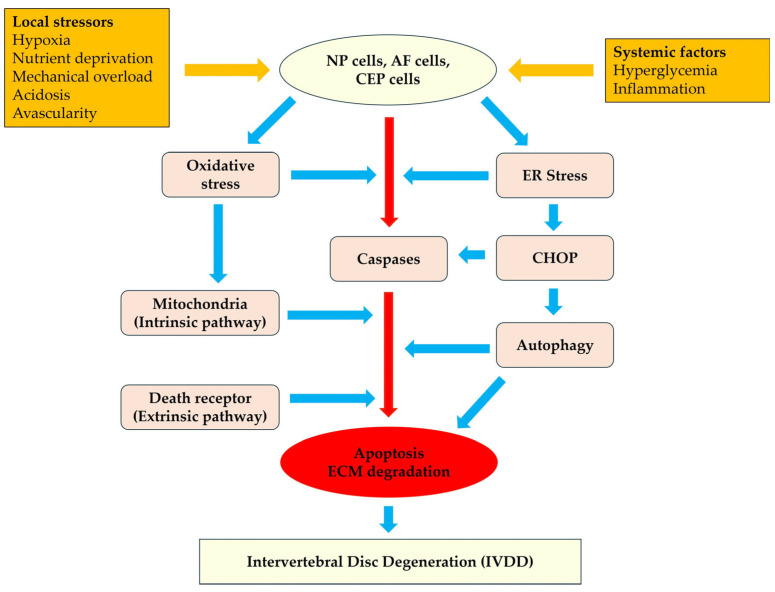
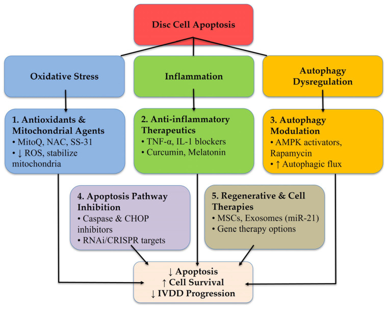
Apoptosis plays a crucial role in the progression of intervertebral disc degeneration (IVDD), a significant cause of chronic low back pain. This review explores disc cell apoptosis's cellular and molecular mechanisms, focusing on nucleus pulposus, annulus fibrosus, and cartilage endplates cells. Apoptotic pathways-intrinsic (mitochondrial), extrinsic (death receptor-mediated), ER stress-mediated, and autophagy-related-are activated by oxidative stress, inflammation, mechanical load, and metabolic disturbances like hyperglycemia. Diabetes exacerbates disc cell apoptosis through AGE-RAGE signaling and mitochondrial dysfunction. Inflammation further amplifies apoptotic cascades via cytokine signaling and ROS generation. The review also examines emerging therapeutic strategies, including antioxidants (e.g., MitoQ, resveratrol), anti-inflammatory agents (e.g., cytokine inhibitors), autophagy modulators (e.g., rapamycin, metformin), and stem cell and gene therapies. While promising preclinical results exist, challenges such as poor bioavailability and clinical translation remain. Enhanced understanding of apoptosis pathways informs future cellular preservation and matrix integrity treatments. Based on a comprehensive literature search from 2000 to 2025, this narrative review synthesizes current knowledge, identifies knowledge gaps, and discusses translational potential. Our findings support a paradigm shift toward mechanism-based therapies that address the root cause of IVDD rather than symptomatic relief alone.
Keywords: ER stress; apoptosis; autophagy; disc cells; hyperglycemia; inflammation; intervertebral disc degeneration; mitochondrial dysfunction.
Conflict of interest statement
The authors declare no conflicts of interest.
Figures
Similar articles
-
Mechanistic Interactions Driving Nucleus Pulposus Cell Senescence in Intervertebral Disc Degeneration: A Multi-Axial Perspective of Mechanical, Immune, and Metabolic Pathways.JOR Spine. 2025 Jul 2;8(3):e70089. doi: 10.1002/jsp2.70089. eCollection 2025 Sep. JOR Spine. 2025. PMID: 40606198 Free PMC article. Review.
-
Intervertebral disc spheroids as anin vitromulticellular platform for recapitulating the microenvironment of intervertebral disc degeneration.Biofabrication. 2025 Jun 26;17(3). doi: 10.1088/1758-5090/ade56c. Biofabrication. 2025. PMID: 40527335
-
RTA 408 attenuates TBHP-Induced apoptosis in nucleus pulposus cells via Nrf2/ARE and NF-κB signaling pathways: in vitro and in vivo evidence for mitigating rats' intervertebral disc degeneration.Arthritis Res Ther. 2025 Jun 19;27(1):128. doi: 10.1186/s13075-025-03588-7. Arthritis Res Ther. 2025. PMID: 40537840 Free PMC article.
-
Exploring the critical role of PANoptosis in the pathogenesis of intervertebral disc degeneration: mechanisms and potential therapeutic targets.Front Cell Dev Biol. 2025 Jun 19;13:1611936. doi: 10.3389/fcell.2025.1611936. eCollection 2025. Front Cell Dev Biol. 2025. PMID: 40612104 Free PMC article. Review.
-
Lentinan attenuates histone deacetylation of SOCS1 promoter to remodel autophagy via JAK2/STAT3 pathway for the prevention of intervertebral disc degeneration.Int J Biol Macromol. 2025 Jul 30;321(Pt 4):146457. doi: 10.1016/j.ijbiomac.2025.146457. Online ahead of print. Int J Biol Macromol. 2025. PMID: 40749910
References
-
- Feng Y., Egan B., Wang J. Overview of intervertebral disc degeneration: Pathophysiology, diagnosis, and treatment. Bone Res. 2022;10:59.
-
- Zhang X.B., Hu Y.C., Cheng P., Zhou H.Y., Chen X.Y., Wu D., Zhang R.H., Yu D.C., Gao X.D., Shi J.T., et al. Targeted therapy for intervertebral disc degeneration: Inhibiting apoptosis is a promising treatment strategy. Int. J. Med. Sci. 2021;18:2799–2813. doi: 10.7150/ijms.59171. - DOI - PMC - PubMed
Publication types
LinkOut - more resources
Full Text Sources
