. 2025 Jun 18;15(12):1555.
doi: 10.3390/diagnostics15121555.

MCM4 as Potential Metastatic Biomarker in Lung Adenocarcinoma

Affiliations

MCM4 as Potential Metastatic Biomarker in Lung Adenocarcinoma

Hung-Chih Lai et al. Diagnostics (Basel). .

Abstract

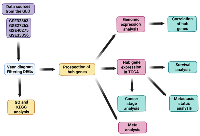
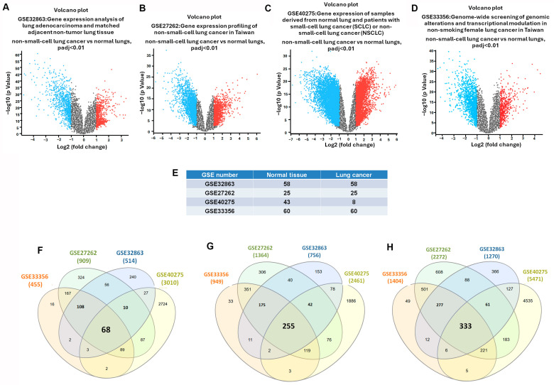
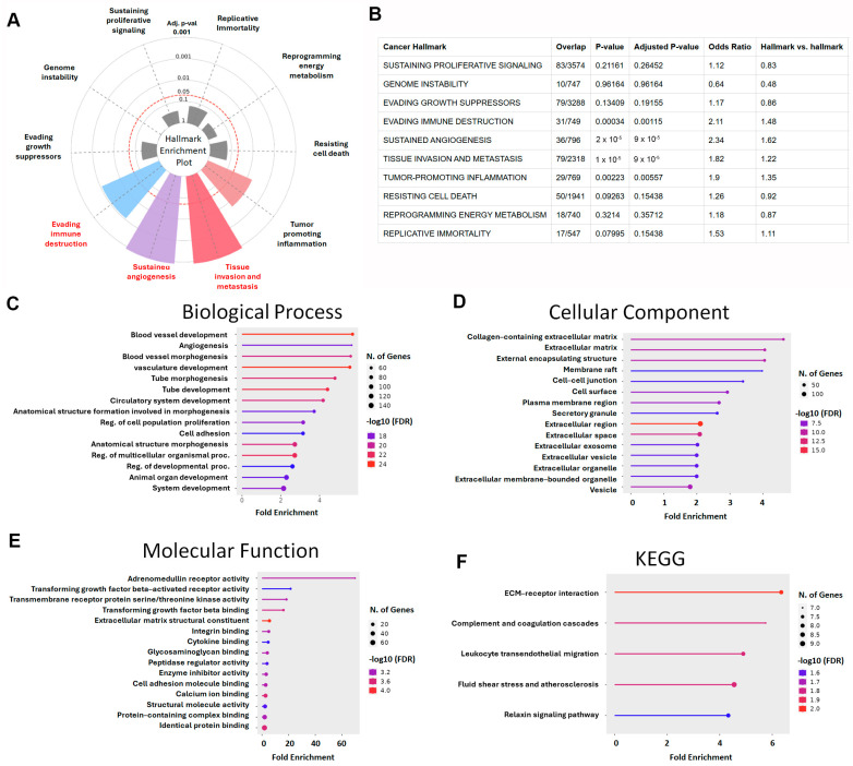
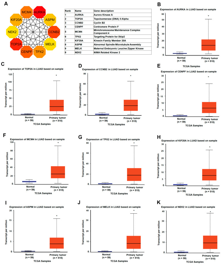
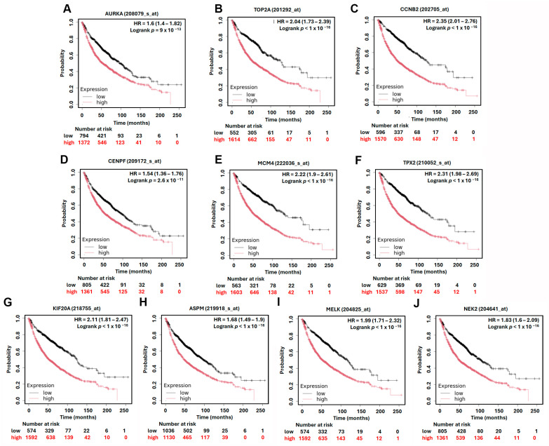
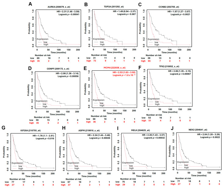
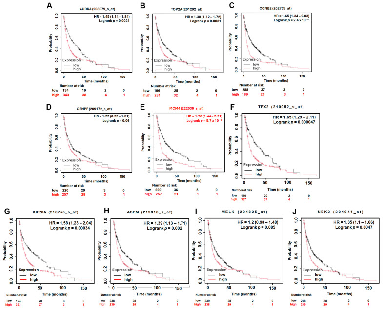
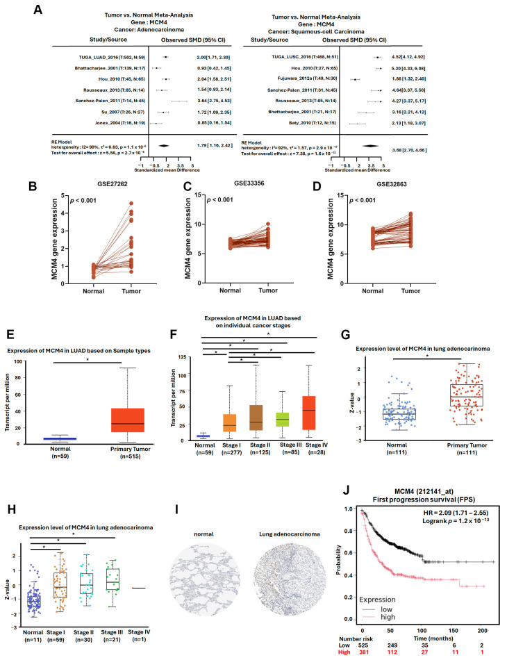
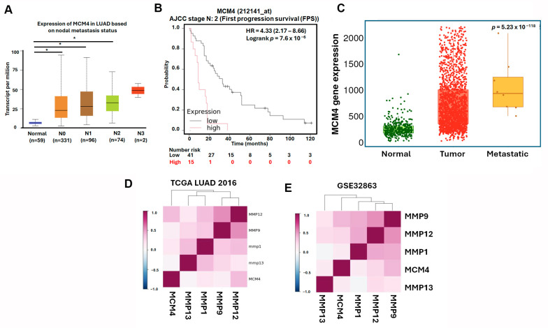
Background: Lung adenocarcinoma (LUAD) is the most common subtype of non-small-cell lung cancer and is frequently diagnosed at advanced stages with metastasis, contributing to its poor prognosis. Identifying key metastasis-related biomarkers is critical for improving early diagnosis and therapeutic targeting. Methods: We analyzed four GEO microarray datasets (GSE32863, GSE27262, GSE40275, and GSE33356) and TCGA data to identify differentially expressed genes (DEGs) in LUAD. Functional enrichment of DEGs was analyzed using Gene Ontology, Kyoto Encyclopedia of Genes and Genomes analysis, and a Cancer Hallmark Enrichment Plot. Hub gene analysis was conducted using Cytoscape. Hub genes were evaluated for their expression, prognostic significance (via the Kaplan-Meier plotter), and clinical correlation using additional platforms (TCGA, Lung Cancer Explorer, TNMplot, and the Human Protein Atlas). Results: A total of 333 consistently dysregulated DEGs were identified, enriched in pathways related to metastasis, including angiogenesis, immune escape, and ECM interaction. Ten hub genes (AURKA, TOP2A, CCNB2, CENPF, MCM4, TPX2, KIF20A, ASPM, MELK, and NEK2) were identified through network analysis. Among these, MCM4 showed strong upregulation in LUAD and was significantly associated with poor overall survival. Notably, MCM4 expression also correlated with post-progression survival and markers of invasiveness. Immunohistochemistry and transcriptomic analyses confirmed MCM4 overexpression at both mRNA and protein levels. Additionally, MCM4 expression was positively correlated with various matrix metalloproteinases, supporting its role in promoting tumor invasiveness. Conclusions:MCM4 is a novel potential biomarker for LUAD metastasis and prognosis. Its consistent upregulation, association with metastatic markers, and clinical significance suggest it may serve as a candidate target for diagnostic use or therapeutic intervention in advanced LUAD.

Keywords: MCM4; differentially expressed genes; lung adenocarcinoma; metastasis.


Conflict of interest statement

The authors declare that they have no conflicts of interest.

Figures

Figure 1
Figure 1
Flowchart of this study.
Figure 2
Figure 2
Intersecting DEGs in GEO datasets of lung cancer: (AD) volcano plots of genes significantly upregulated (red) and downregulated (blue) in lung tumors compared to normal tissue; (E) sample sizes: GSE32863 (58 normal and 58 lung cancer tissues), GSE27262 (25 normal and 25 lung cancer tissues), GSE40275 (43 normal and 8 lung cancer tissues), and GSE33356 (60 normal and 60 lung cancer tissues); (FH) Venn diagrams of overlapping upregulated, downregulated, and all DEGs.
Figure 3
Figure 3
Functional enrichment analysis of LUAD-associated DEGs: (A) Hallmark Enrichment Plot displaying the distribution of DEGs across ten canonical cancer hallmarks. Colored slices represent significantly enriched hallmarks (adjusted p < 0.05), and slice size indicates enrichment strength. (B) This table summarizes the enrichment of LUAD-associated differentially expressed genes (DEGs) across ten classical cancer hallmarks using the Cancer Hallmarks Analytics Tool. Columns include cancer hallmark, overlap (DEGs/reference), p-value, adjusted p-value, odds ratio, and hallmark vs. hallmark ratio. (C) Gene Ontology (GO) biological process enrichment analysis of DEGs. (D) GO cellular component enrichment analysis. (E) GO molecular function enrichment analysis. (F) KEGG pathway enrichment analysis of DEGs (color marks false discovery rate).
Figure 4
Figure 4
Identification and expression analysis of LUAD-associated hub genes: (A) the top 10 hub genes were identified using the Maximal Clique Centrality (MCC) algorithm in the cytoHubba plugin within Cytoscape; (BK) gene expression analysis of the top 10 hub genes (AURKA, TOP2A, CCNB2, TPX2, MCM4, KIF20A, CENPF, ASPM, NUSAP1, and NEK2) in lung adenocarcinoma (LUAD) and normal lung tissues based on TCGA RNA-seq data. * p-value < 0.05.
Figure 5
Figure 5
Kaplan–Meier overall survival analysis of LUAD patients based on the expression of top 10 hub genes. High and low expression groups are represented by red and black lines, respectively. Hazard ratios (HRs) and log-rank p-values are indicated in each plot. (A) AURKA, (B) TOP2A, (C) CCNB2, (D) CENPF, (E) MCM4, (F) TPX2, (G) KIF20A, (H) ASPM, (I) MELK, (J) NEK2.
Figure 6
Figure 6
Kaplan–Meier overall survival in AJCC N2 subtype analysis of LUAD patients based on the expression of top 10 hub genes. High and low expression groups are represented by red and black lines, respectively. Hazard ratios (HRs) and log-rank p-values are indicated in each plot. (A) AURKA, (B) TOP2A, (C) CCNB2, (D) CENPF, (E) MCM4, (F) TPX2, (G) KIF20A, (H) ASPM, (I) MELK, (J) NEK2.
Figure 7
Figure 7
Kaplan–Meier post-progression survival (PPS) analysis of LUAD patients based on the expression of top 10 hub genes. High and low expression groups are represented by red and black lines, respectively. Hazard ratios (HRs) and log-rank p-values are indicated in each plot. (A) AURKA, (B) TOP2A, (C) CCNB2, (D) CENPF, (E) MCM4, (F) TPX2, (G) KIF20A, (H) ASPM, (I) MELK, (J) NEK2.
Figure 8
Figure 8
MCM4 is highly expressed in LUAD and correlates with tumor progression and early poor prognosis: (A) meta-analysis of MCM4 expression in LUAD and LUSC compared to normal lung tissues, obtained from the Lung Cancer Explorer (LCE); (BD) paired t-test analysis of MCM4 expression in tumor and adjacent normal tissues from GEO datasets GSE32863, GSE33356, and GSE27262; (E,F) MCM4 expression in LUAD across different clinical stages (stage I–IV) using TCGA data; (G,H) MCM4 protein expression in LUAD and normal tissues based on TCGA proteomic analysis; (I) immunohistochemistry (IHC) staining results from the Human Protein Atlas demonstrating MCM4 protein expression in LUAD and normal lung tissues; (J) KM plot showing First Progression Survival (FPS) in LUAD patients stratified by MCM4 expression level. * p-value < 0.05.
Figure 9
Figure 9
MCM4 expression is associated with lymph node metastasis and adverse survival outcomes in advanced LUAD: (A) MCM4 mRNA expression levels in LUAD samples stratified by lymph node metastasis status; (B) KM plot of (First Progression Survival (FPS) in LUAD patients with AJCC N2 stage; and (C) MCM4 expression analysis from the TNMplot platform across normal lung, primary LUAD, and metastatic LUAD tissues. Statistical significance was assessed using the Kruskal–Wallis test. (D,E) Correlation analysis of MCM4 and matrix metalloproteinases (MMP1, MMP9, MMP12, and MMP13) expression levels in LUAD using Lung Cancer Explorer. * p-value < 0.05.

Similar articles

References

    1. Sung H., Ferlay J., Siegel R.L., Laversanne M., Soerjomataram I., Jemal A., Bray F. Global Cancer Statistics 2020: GLOBOCAN Estimates of Incidence and Mortality Worldwide for 36 Cancers in 185 Countries. CA Cancer J. Clin. 2021;71:209–249. doi: 10.3322/caac.21660. - DOI - PubMed
    1. Molina J.R., Yang P., Cassivi S.D., Schild S.E., Adjei A.A. Non-small cell lung cancer: Epidemiology, risk factors, treatment, and survivorship. Mayo Clin. Proc. 2008;83:584–594. doi: 10.1016/S0025-6196(11)60735-0. - DOI - PMC - PubMed
    1. Horvath L., Thienpont B., Zhao L., Wolf D., Pircher A. Overcoming immunotherapy resistance in non-small cell lung cancer (NSCLC)—Novel approaches and future outlook. Mol. Cancer. 2020;19:141. doi: 10.1186/s12943-020-01260-z. - DOI - PMC - PubMed
    1. Lahiri A., Maji A., Potdar P.D., Singh N., Parikh P., Bisht B., Mukherjee A., Paul M.K. Lung cancer immunotherapy: Progress, pitfalls, and promises. Mol. Cancer. 2023;22:40. doi: 10.1186/s12943-023-01740-y. - DOI - PMC - PubMed
    1. Hanahan D. Hallmarks of Cancer: New Dimensions. Cancer Discov. 2022;12:31–46. doi: 10.1158/2159-8290.CD-21-1059. - DOI - PubMed

LinkOut - more resources