Review
. 2025 Jun 6;26(12):5441.
doi: 10.3390/ijms26125441.

The Role of the RecFOR Complex in Genome Stability

Affiliations
Review

The Role of the RecFOR Complex in Genome Stability

Piero R Bianco. Int J Mol Sci. .

Abstract

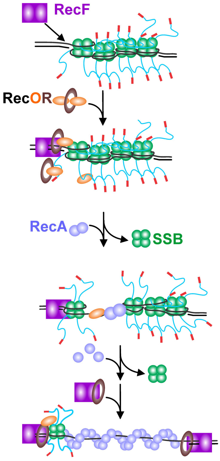
The maintenance of genome stability requires the coordinated actions of multiple proteins and protein complexes. One critical family of proteins is the recombination mediators. Their role is to facilitate the formation of recombinase nucleoprotein filaments on single-stranded DNA (ssDNA). Filament formation can take place on post-replicative ssDNA gaps as well as on 3'-tailed duplexes resulting from helicase-nuclease processing. In prokaryotes, the RecF, O, and R proteins are widely distributed and mediate RecA loading as either the RecFOR or RecOR complexes, depending on the species being studied. In this review, I compare and contrast the available biochemical and structural information to provide insight into the mechanism of action of this critical family of mediators.

Keywords: DNA repair; RecF; RecO; RecR; SSB protein; genetic recombination; recombination mediator.


Conflict of interest statement

The author declares no conflict of interest.

Figures

Figure 4
Figure 4
RecO’s structural features are conserved. (A) The RecO protein from Campylobacter jejuni (PDB file:7 YMO). The three domains of RecO are indicated. The image was generated using UCSF Chimera [85]. (B) An SSB C-terminal peptide binds in the α-helical domain. The structure of the E.coli RecO protein bound to a peptide corresponding to the last eight residues is shown. PDB File 3 Q8 D was used to generate the image using Discovery Studio (Biovia). The Connolly surfaces for the OB-fold (green), C-terminal domain (mauve), and peptide (red) are shown [77]. (C,D) The E.coli RecO OB-fold binds to the intrinsically disordered linker of its cognate SSB protein. The OB-fold from 3 Q8 D was aligned with the SH3 domains of PDB files insulin receptor tyrosine kinase substrate 2 XKC (C) and abl tyrosine kinase 1 ABO (D), respectively [49]. The Connolly surfaces for the OB-fold (green), C-terminal domain (mauve), and PXXP–ligands (red and orange, respectively) are shown [77]. In panel (C), the OB-fold/ligand interaction is viewed from the top as in panel (B). In (D), the RecO–PXXP–ligand complex is viewed from the side.
Figure 1
Figure 1
Mechanisms of RecA filament nucleation. (A) RecBCD loads RecA in a chi-dependent manner. Once the translocating RecBCD encounters an appropriately oriented chi-sequence (χEc; chi = χ = crossover hotspot instigator), it pauses, and the nuclease activity on the 3′-terminated strand is attenuated while the nuclease activity on the 5′-terminated strand is upregulated. Concurrently, the RecA-loading domain is exposed so that RecA protein can be specifically loaded onto the now intact 3′-terminated strand once the χ-modified enzyme continues translocating and unwinding DNA past the chi sequence. (B) In the absence of RecBCD, RecQ helicase unwinds the DNA duplex while RecJ degrades the 5′-terminated strand. SSB binds to the exposed 3′-terminated single strand of DNA. RecFOR binds to the DNA (possibly at the ss–dsDNA junction) and RecOR mediates the displacement of SSB concomitant with the loading of RecA. (C) The ssDNA in post-replicative gaps is bound by SSB. RecFOR binds to the DNA (possibly at the ss–dsDNA junction) and RecOR mediates displacement of SSB concomitant with the nucleation of an RecA filament. RecFR limits the extension of the RecA filament into the duplex DNA region downstream of the gap. (D) In organisms other than E. coli, AddAB (or AdnAB) unwinds duplex DNA from an exposed end and simultaneously degrades the 5′-terminated strand, leaving the 3′-terminated strand intact, which is bound by SSB. These helicase/nucleases are not known to load RecA, which is instead facilitated by RecFOR, concomitant with the displacement of SSB.
Figure 2
Figure 2
RecF, O, and R protein domain organizations are shared among different bacteria. The schematics for each protein are adapted from [50]. Proteins are presented in order of decreasing size, with the relevant domains color-coded. The regions of each protein responsible for mediating interactions with partners are presented below each schematic. Details are discussed in the text.
Figure 3
Figure 3
The RecF, O, and R proteins assemble into distinct functional, higher-order structures. Images were generated using Discovery Studio (Biovia). The Connolly surfaces for each protomer are shown [77]. (A) RecR can assemble into a tetramer. The four subunits (each shown in a different color) assemble to form a ring with a central hole sufficiently large to accommodate dsDNA. (B) The RecOR complex is shown from the side. Two subunits of the RecR tetramer are seen in this view with two RecO monomers bound. Each RecO binds to RecR via its N-terminal OB-fold (colored green). The remaining residues of each ReO are colored mauve to facilitate visualization. (C). The RecFOR complex bound to dsDNA is viewed from two angles. Left: the complex is viewed from the side with all protomers of RecF colored blue and RecR colored orange. The N-terminal OB-fold of RecO is colored green, identical to panel (B), and is bound to RecR in proximity to the duplex DNA end. Right: the complex is rotated to enable visualization of the interaction of RecF with the DNA. From this angle, the RecF dimer straddles the duplex DNA, anchoring the complex.
Figure 5
Figure 5
General scheme for RecFOR-mediated loading of RecA onto SSB-coated ssDNA gaps. In the scheme shown, SSB binds to and coats an ssDNA gap. In the first step of RecA loading, the RecF dimer binds to the ss–dsDNA junction. This directs loading of RecOR as either the RecR4–RecO2 or RecR4–RecO complex onto the DNA. This positions RecO in proximity of SSB. The binding of the SSB linker and/or tip to RecO leads to SSB repositioning or dissociation, thereby exposing the ssDNA. RecO may dissociate along with SSB or remain bound to the ssDNA, leaving a RecFR complex at the proximal ss–dsDNA junction. RecA dimers bind to the exposed ssDNA, thereby causing nucleating filament formation. Additional RecA binds extend the filament to coat the ssDNA gap. To limit filament extension into the downstream duplex DNA, a second RecFR complex binds to the distal end of the filament or the ss–dsDNA junction.

Similar articles

References

    1. Bianco P.R., Tracy R.B., Kowalczykowski S.C. DNA strand exchange proteins: A biochemical and physical comparison. Front. Biosci. 1998;3:D570–D603. - PubMed
    1. Bianco P.R. eLS. John Wiley & Sons, Ltd.; Hoboken, NJ, USA: 2001. RecA Protein; pp. 1–12.
    1. Roman L.J., Dixon D.A., Kowalczykowski S.C. RecBCD-dependent joint molecule formation promoted by the Escherichia coli RecA and SSB proteins. Proc. Natl. Acad. Sci. USA. 1991;88:3367–3371. doi: 10.1073/pnas.88.8.3367. - DOI - PMC - PubMed
    1. Dillingham M.S., Kowalczykowski S.C. RecBCD enzyme and the repair of double-stranded DNA breaks. Microbiol. Mol. Biol. Rev. 2008;72:642–671. doi: 10.1128/MMBR.00020-08. - DOI - PMC - PubMed
    1. Wigley D.B. Bacterial DNA repair: Recent insights into the mechanism of RecBCD, AddAB and AdnAB. Nat. Rev. Microbiol. 2013;11:9–13. doi: 10.1038/nrmicro2917. - DOI - PubMed

MeSH terms

LinkOut - more resources