. 2025 Jun 6;26(12):5457.
doi: 10.3390/ijms26125457.

Electrospun DegraPol Tube Delivering Stem Cell/Tenocyte Co-Culture-Derived Secretome to Transected Rabbit Achilles Tendon-In Vitro and In Vivo Evaluation

Affiliations

Electrospun DegraPol Tube Delivering Stem Cell/Tenocyte Co-Culture-Derived Secretome to Transected Rabbit Achilles Tendon-In Vitro and In Vivo Evaluation

Julia Rieber et al. Int J Mol Sci. .

Abstract

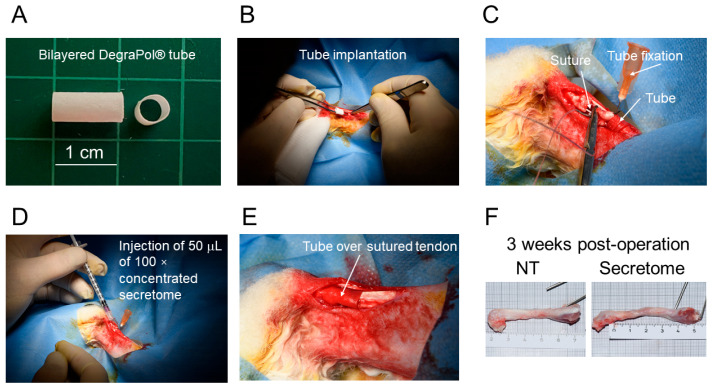
Tendon ruptures have recently reached incidences of 18-35 cases/100,000 and often lead to adhesion formation during healing. Furthermore, scar formation may result in inferior biomechanics and often leads to re-ruptures. To address these problems, we cultivated rabbit adipose-derived stem cells in a co-culture with rabbit Achilles tenocytes and harvested their secretome. Following a cell-free approach, we incorporated such secretome into an electrospun tube via emulsion electrospinning. These novel implants were characterized by SEM, the WCA, and FTIR. Then, they were implanted in the rabbit Achilles tendon full transection model with an additional injection of secretome, and the adhesion extent as well as the biomechanics of extracted tendons were assessed three weeks postoperatively. The fiber thickness was around 3-5 μm, the pore size 11-13 μm, and the tube wall thickness approximately 265 μm. The WCA indicated slightly hydrophilic surfaces in the secretome-containing layer, with values of 80-90°. In vivo experiments revealed a significant reduction in adhesion formation (-22%) when secretome-treated tendons were compared to DegraPol® (DP) tube-treated tendons (no secretome). Furthermore, the cross-sectional area was significantly smaller in secretome-treated tendons compared to DP tube-treated ones (-32%). The peak load and stiffness of secretome-treated tendons were not significantly different from native tendons, while tendons treated with pure DP tubes exhibited significantly lower values. We concluded that secretome treatment supports tendon healing, with anti-adhesion effects and improved biomechanics at 3 weeks, making this approach interesting for clinical application.

Keywords: adhesion; biomechanics; mesenchymal stem cells; rabbit; secretome; tendon healing; tenocytes.


Conflict of interest statement

The authors declare no conflicts of interest.

Figures

Figure 1
Figure 1
An overview of the experiments conducted in this study. First, adipose-derived stem cells and Achilles tenocytes were harvested from New Zealand White rabbits (1). Second, a co-culture of rabbit ASCs and rabbit tenocytes in a ratio of 3:1 was used to collect the secretome (indicated by a scheme of colored triangles and rectangles) (2). Third, after concentrating this secretome, a water-in-oil emulsion was made where the secretome was in the aqueous phase and the polymer DegraPol® was in the chloroform/HFP phase. This emulsion was electrospun to yield a bilayer tubular implant material that was characterized by SEM, the WCA, and FTIR (3). Fourth, the tube was implanted in a rabbit full-transection model. Upon harvest, the biomechanics of the Achilles tendons and their adhesion extent were assessed three weeks post-operation (4).
Figure 2
Figure 2
SEM images of the inner and outer surface of electrospun pure DegraPol fiber meshes (DP = control) or secretome/DP meshes (Secretome); (A) fiber thickness (B) and pore size (C). Data is shown as box-and-whisker plots, showing the interquartile range and the full data range. The normality was assessed using the Shapiro–Wilk test. As data were normally distributed, group comparisons were performed using a one-way ANOVA followed by Tukey’s multiple comparisons test (DP, n = 9, secretome, n = 6). p-values ≤ 0.05 were considered significant and are denoted as follows: * p ≤ 0.05; *** p ≤ 0.001, and **** p ≤ 0.0001.
Figure 3
Figure 3
Static (A) and dynamic water contact angles (B) of pure DP meshes (=control) and of emulsion electrospun secretome/DP fiber meshes. Data is shown as box-and-whisker plots, showing the interquartile range and the full data range. The normality was assessed using the Shapiro–Wilk test. As data were normally distributed, group comparisons were performed using a one-way ANOVA followed by Tukey’s multiple comparisons test. For DP, n = 9; for secretome, n = 6 (A). Data is shown as the mean and SD. The Kruskal–Wallis test with Dunn’s multiple comparison was used for comparisons of advancing, receding angle, and hysteresis (B). p-values ≤ 0.05 were considered significant and are denoted as ** p ≤ 0.01.
Figure 4
Figure 4
FTIR spectra of three pure DP tubes (Control) and two secretome/DP tubes (Secretome) (A) and the calculated C=O-to-C–O ratio of intensities for the two materials (B). Data is shown as the mean and SD. The Mann–Whitney test was used for comparisons of the C=O-to-C–O ratio (B). No significant difference could be determined.
Figure 5
Figure 5
The tube before implantation (A). After flipping the tube inside-out to provide the secretome layer towards the tendon, it was pulled over the proximal stump of the fully transected Achilles tendon (B). To avoid back slippage, the tube was fixed in position by a cannula during suturing (C). A volume of 50 μL of 100× concentrated secretome was injected after finishing the suture (D). After the removal of the cannula, the tube was pulled over the sutured tendon (E). Three weeks post-operation, the extracted tendons of secretome-treated and contralateral non-treated (NT) tendons appeared similar during macroscopic inspection (F).
Figure 6
Figure 6
From histological cross-sections, the adhesion extent was assessed as a fraction by measuring the adherent distances and dividing the sum of them by the whole circumference, exemplified with H&E-stained sections (A). The fraction of adherent tissue is presented for the four experimental groups; non-treated contralateral legs that did not have any intervention (NT), repaired tendons with a pure DP tube (DP), repaired tendons with a secretome/DP tube (secretome), and repaired tendons without a tube (4-strand, indicating the type of suture used to repair the tendon) (B). Data is shown as the mean and SD. The normality was assessed using the Shapiro–Wilk test. As the data were normally distributed, group comparisons were performed using a one-way ANOVA followed by Fisher’s PLSD multiple comparisons test. (n = 11 for 4-strand, DP and secretome, n = 61 for NT [20]). p-values ≤ 0.05 were considered significant and are denoted as follows: * p ≤ 0.05, *** p ≤ 0.001.
Figure 7
Figure 7
The biomechanical properties of the repaired tendons, either treated with a pure DP tube (DP) or with a secretome tube and a secretome injection (Secretome), compared to healthy non-treated tendons (NT). Data is shown as the mean and SD for length (A), cross sectional area (CSA) (B), load until failure (C), elastic modulus (D), stiffness (E), and failure stress (F). The groups were compared using the nonparametric Kruskal–Wallis test for the length, elastic modulus, and stiffness (no normal distribution of data). As data were normally distributed, groups of the following parameters were compared using a one-way ANOVA: CSA, load until failure, and failure stress. p-values ≤ 0.05 were considered significant and are denoted as follows: * p ≤ 0.05; ** p ≤ 0.01, *** p ≤ 0.001, **** p ≤ 0.0001; not significant (ns).

Similar articles

References

    1. Liang W., Zhou C., Deng Y., Fu L., Zhao J., Long H., Ming W., Shang J., Zeng B. The current status of various preclinical therapeutic approaches for tendon repair. Ann. Med. 2024;56:2337871. doi: 10.1080/07853890.2024.2337871. - DOI - PMC - PubMed
    1. Hoeffner R., Agergaard A.S., Svensson R.B., Cullum C., Mikkelsen R.K., Konradsen L., Krogsgaard M., Boesen M., Kjaer M., Magnusson S.P. Tendon Elongation and Function After Delayed or Standard Loading of Surgically Repaired Achilles Tendon Ruptures: A Randomized Controlled Trial. Am. J. Sports Med. 2024;52:1022–1031. doi: 10.1177/03635465241227178. - DOI - PubMed
    1. Schulze-Tanzil G.G., Delgado-Calcares M., Stange R., Wildemann B., Docheva D. Tendon healing: A concise review on cellular and molecular mechanisms with a particular focus on the Achilles tendon. Bone Jt. Res. 2022;11:561–574. doi: 10.1302/2046-3758.118.BJR-2021-0576.R1. - DOI - PMC - PubMed
    1. Darrieutort-Laffite C., Blanchard F., Soslowsky L.J., Le Goff B. Biology and physiology of tendon healing. Jt. Bone Spine. 2024;91:105696. doi: 10.1016/j.jbspin.2024.105696. - DOI - PubMed
    1. Sharma P., Maffulli N. Tendon injury and tendinopathy: Healing and repair. J. Bone Jt. Surg. Am. 2005;87:187–202. doi: 10.2106/JBJS.D.01850. - DOI - PubMed

LinkOut - more resources